. 2020 Dec 23;10(1):22373.
doi: 10.1038/s41598-020-79450-4.

Schistosoma mansoni eggs induce Wnt/β-catenin signaling and activate the protooncogene c-Jun in human and hamster colon

Affiliations

Schistosoma mansoni eggs induce Wnt/β-catenin signaling and activate the protooncogene c-Jun in human and hamster colon

Jakob Weglage et al. Sci Rep. .

Abstract

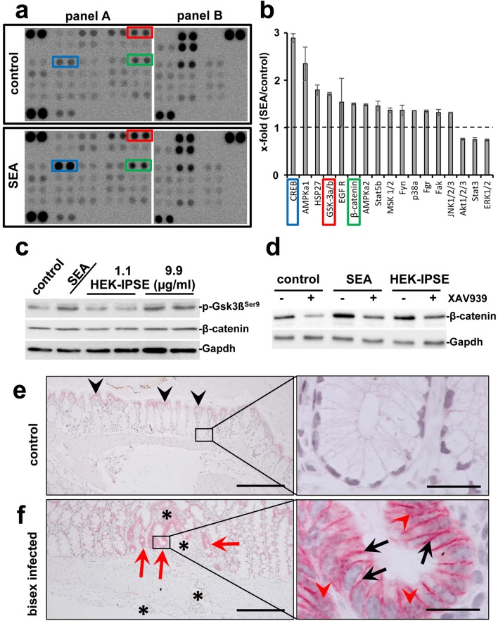
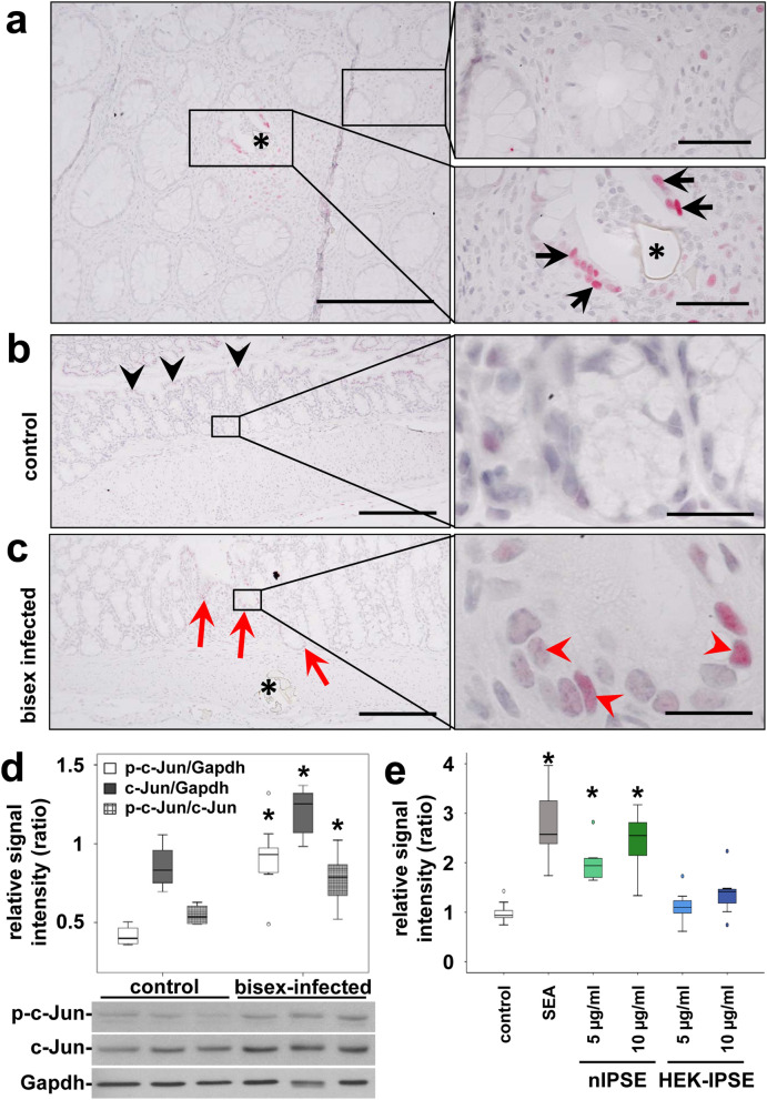
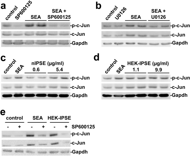
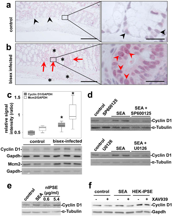
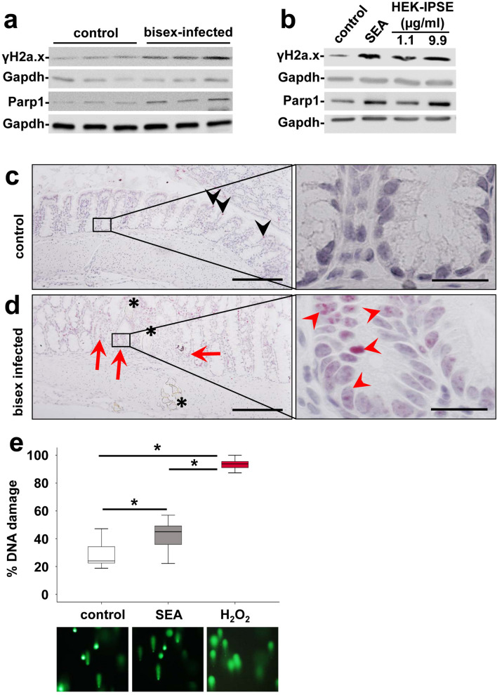
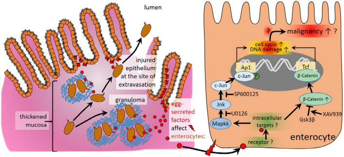
Schistosomiasis (bilharzia) is a neglected tropical disease caused by parasitic flatworms of the genus Schistosoma, with considerable morbidity in parts of the Middle East, South America, Southeast Asia, in sub-Saharan Africa, and particularly also in Europe. The WHO describes an increasing global health burden with more than 290 million people threatened by the disease and a potential to spread into regions with temperate climates like Corsica, France. The aim of our study was to investigate the influence of S. mansoni infection on colorectal carcinogenic signaling pathways in vivo and in vitro. S. mansoni infection, soluble egg antigens (SEA) and the Interleukin-4-inducing principle from S. mansoni eggs induce Wnt/β-catenin signaling and the protooncogene c-Jun as well as downstream factor Cyclin D1 and markers for DNA-damage, such as Parp1 and γH2a.x in enterocytes. The presence of these characteristic hallmarks of colorectal carcinogenesis was confirmed in colon biopsies from S. mansoni-infected patients demonstrating the clinical relevance of our findings. For the first time it was shown that S. mansoni SEA may be involved in the induction of colorectal carcinoma-associated signaling pathways.


Conflict of interest statement

The authors declare no competing interests.

Figures

Figure 1
Figure 1
Eggs of S. mansoni induce Wnt-signaling/ß-Catenin in colon epithelium. (a) Phosphokinase proteome profiler arrays and (b) densitometric assessment thereof demonstrated the induction of β-catenin as well as the phosphorylation of Gsk3β and CREB in SW620 cells after soluble egg antigen (SEA)-stimulation. The 17 most regulated factors of the array are depicted in (b). The Human Phospho-Kinase Array is a tool to simultaneously detect the relative levels of phosphorylation of kinases and two related total proteins. The array consists of two membranes (left and right membrane of each panel) on which each kinase or substrate was analyzed in duplicates. The experiment and assay was performed twice. The positions of β-catenin (green box), pGsk3β (red box), and CREB (blue box) are highlighted. (c) Western blot analysis showed the phosphorylation of Gsk3β and indicated a slightly enhanced β-catenin expression in SW620 cells stimulated with SEA and HEK-IPSE for 4 h. Gapdh was used as loading control. The experiment and assay were reproduced at least two times. Representative blots are shown. The quantification is presented in Suppl. Fig. 1. (d) Inhibition of Wnt-signaling by the tankyrase inhibitor XAV939 demonstrated a reduction of SEA-induced β-catenin. The experiment and assay were reproduced at least two times. The cells were pretreated with XAV939 for 15 h and during the 4-h-stimulation. Representative blots are shown. The quantification is presented in Suppl. Fig. 2. (e) Immunostaining of β-catenin in the colon of control hamsters visualized low amounts of epithelial β-catenin at the luminal side of the colon (black arrowheads), but not in those enterocytes inside the crypts (magnification of the boxed area is depicted in the right panel). (f) Enhanced expression of β-catenin was observed at the bottom of those crypts around the extravasation sites of S. mansoni eggs in bisex-infected hamster colon (red arrows). Please note that enhanced nuclear translocation of β-catenin in enterocytes at the bottom of those crypts, which surround the extravasation sites of S. mansoni eggs in bisex-infected hamster colon (red arrowheads in the magnified area, right panel). The black arrows depict enhanced intercellular accumulation of β-catenin. S. mansoni eggs (*), magnification 200×, bar 200 µm (left panels) and 1000×, bar 20 µm (right panels). The quantification is presented in Suppl. Fig. 3.
Figure 2
Figure 2
Activation of the protooncogene c-Jun in enterocytes at extravasation sites of S. mansoni eggs. (a) A representative histologic slice of a rectal biopsy from an 29 year-old Egyptian male with schistosomal colitis is shown. Immunohistochemical staining of c-Jun (red) depicted its nuclear translocation (arrows) in enterocytes of crypts in direct vicinity of S. mansoni eggs (*, lower right panel). Nearly no nuclear translocation of c-Jun was observed in enterocytes lining unaffected crypts at a distance of at least 200 µm (upper right panel). The same result was shown in at least three samples from different patients. Co-staining of nuclei in blue, magnification 200× and 1000×, bars 200 µm (left panel) and 50 µm (panel on the right). (b) Immunostaining visualized low amounts of epithelial c-Jun at the luminal side of the colon (black arrowheads), but not in enterocytes inside the crypts (magnification of the boxed area is depicted in the right panel). (c) Immunostaining demonstrated enhanced nuclear translocation of c-Jun (red arrowheads) in enterocytes of crypts (red arrows) adjacent to sites of S. mansoni egg (*) extravasation in bisex-infected hamsters. The quantification is presented in Suppl. Fig. 5. Magnified representative crypts are shown on the right. Magnification in (b,c) 200× and 1000×, bars 200 µm and 20 µm. (d) Western blot analysis and subsequent semi-quantitative assessment of optical density suggested an enhanced expression and activation of c-Jun (p-Ser73) in the colon of S. mansoni bisex-infected hamsters in comparison to single sex-infected hamsters, lacking egg production. The experiment and assay were reproduced at least two times. Representative blots are shown, control n = 4, bisex infection n = 11. Differences were analyzed statistically between control and bisex-infected for the activated form (p–c-Jun), total expression (c-Jun), and the ratio between p–c-Jun and c-Jun. *p ≤ 0.05. (e) The reporter gene assay demonstrated, that SEA and IPSE stimulation lead to the functional activation of the AP1 promotor. The experiment and assay were reproduced at least two times and analyzed by Kruskall-Wallis test. *p ≤ 0.05.
Figure 3
Figure 3
SEA and IPSE activated c-Jun in vitro. (a,b) The expression and phosphorylation of c-Jun increased following SEA-stimulation and was inhibited by additional treatment with the JNK-inhibitor SP600125 (a) and the MEK-inhibitor U0126 (b) in SW620 cells. (c,d) A concentration-dependent induction of expression and activation of c-Jun by nIPSE (c) and HEK-IPSE (d) was shown by western blot. (e) SEA- and HEK-IPSE-induced activation of c-Jun was inhibited by the JNK-inhibitor SP600125. The experiment and assay were reproduced at least two times. Representative blots are shown. The cells were pretreated with inhibitors for 30 min and subsequently for 4 h during stimulation.
Figure 4
Figure 4
S. mansoni egg-induced expression of proliferation markers. (a,b) Increased enterocyte proliferation, denoted by Cyclin D1-immunostaining adjacent [red arrowheads in (b)] to the eggs (*) was distinct from physiological proliferation of enterocytes in basal crypts [black arrowheads in (a)]. Representative cross sections of colon mucosa from hamsters are shown.(a) single sex-infected control, (b) bisex-infected. Magnification 200×, bar 2200 µm and 1000×, bar 20 µm. (c) Western blot analysis and subsequent semi-quantitative assessment of optical density suggested an enhanced expression of Cyclin D1 and Mcm2 in the colon of S. mansoni bisex-infected hamsters in comparison to single sex-infected controls. Representative blots are shown, control n = 4, bisex infection n = 11. Differences were analyzed statistically between control and bisex-infected for Cyclin D1/Gapdh and Mcm2/Gapdh. *p ≤ 0.05. (d) The expression of Cyclin D1 was induced by SEA and could be inhibited by additional treatment with the JNK-inhibitor SP600125 (upper blots) and the MEK-inhibitor U0126 (lower blots) in SW620 cells. The cells were pretreated with inhibitors for 30 min and subsequently during stimulation for 4 h. (e) The expression of Cyclin D1 was also induced by nIPSE. (f) SEA-stimulated Cyclin D1 expression was reduced by tankyrase inhibition, using XAV939, in SW620 cells. IPSE-induced Cyclin D1 expression, however, was not reduced by tankyrase inhibition. The cells were pretreated with XAV939 for 15 h and subsequently during stimulation for 4 h. The experiment and assay was reproduced at least two times. Representative western blots are shown.
Figure 5
Figure 5
S. mansoni egg-induced DNA repair. (a) Western blot analysis demonstrated enhanced expression of γH2a.x and Parp-1 in the colon of S. mansoni bisex-infected hamsters in comparison to single sex-infected controls. Representative blots are shown. (b) Induction of expression and activation of γH2a.x and Parp1 by SEA and HEK-IPSE is shown by western blot (stimulation 4 h). (c,d) Immunostaining of γH2a.x (blue) depicted nuclear staining (red arrows and arrowheads) in enterocytes of crypts in direct vicinity of S. mansoni eggs (*). Representative histologic section of the colon of a control hamster (c) and a bisex-infected hamster (d) are shown. Quantification of nuclear γH2a.x is demonstrated in Suppl. Fig. 10. Nuclear staining of γH2a.x in red, magnification 200×, bar 200 µm and 1000×, bar 20 µm. (e) The comet assay demonstrated enhanced DNA damage by SEA stimulation in SW620 cells. western blot.
Figure 6
Figure 6
Schematic summary of the main findings of the current study. Granuloma formation around S. mansoni eggs trapped within colon tissue is a hallmark of schistosomal colitis. The eggs are metabolically active and highly antigenic. Egg secreted factors (red circles) activate CRC-associated signaling pathways (JNK/c-Jun, Wnt/β-catenin), which initiate proliferation and DNA-damage. These biomolecular processes upon the exposure to schistosomiasis may characterize underlying mechanisms for the predisposition to the development of colorectal cancer as suggested recently.

References

    1. Schistosomiasis—fact sheet. https://www.who.int/news-room/fact-sheets/detail/schistosomiasis (2019).
    1. Schwartz C, Fallon PG. Schistosoma, "eggs-iting" the host: granuloma formation and egg excretion. Front. Immunol. 2018;9:2492. doi: 10.3389/fimmu.2018.02492. - DOI - PMC - PubMed
    1. Hatz CFR. Schistosomiasis. An underestimated problem in industrialized countries? J. Travel Med. 2005;12:1–2. doi: 10.2310/7060.2005.00001. - DOI - PubMed
    1. Lingscheid T, et al. Schistosomiasis in European travelers and migrants. analysis of 14 years TropNet surveillance data. Am. J. Trop. Med. Hyg. 2017;97:567–574. doi: 10.4269/ajtmh.17-0034. - DOI - PMC - PubMed
    1. Boissier J, et al. Outbreak of urogenital schistosomiasis in Corsica (France). An epidemiological case study. Lancet Infect. Dis. 2016;16:971–979. doi: 10.1016/S1473-3099(16)00175-4. - DOI - PubMed

Publication types