. 2020 Dec 23;15(12):e0244215.
doi: 10.1371/journal.pone.0244215. eCollection 2020.

Detailed genetic and functional analysis of the hDMDdel52/mdx mouse model

Affiliations

Detailed genetic and functional analysis of the hDMDdel52/mdx mouse model

Alper Yavas et al. PLoS One. .

Abstract

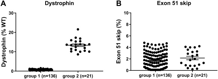
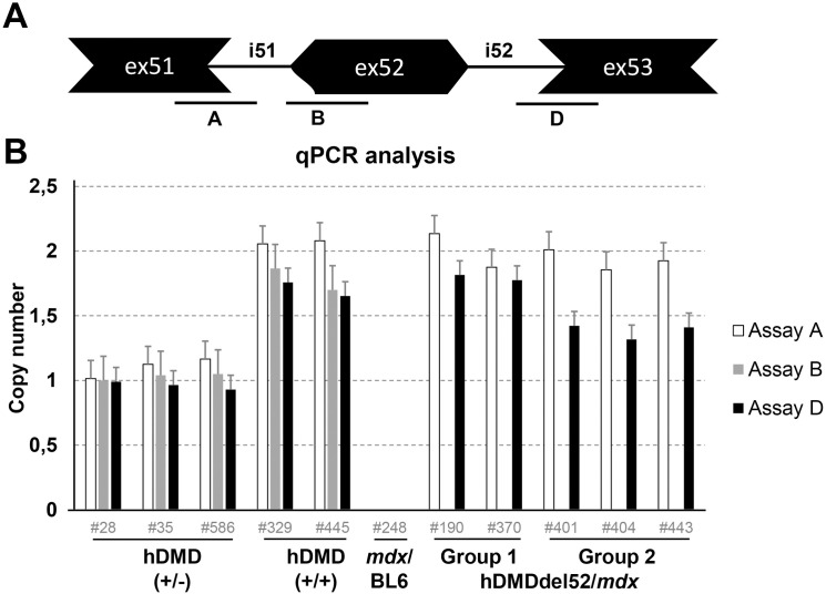
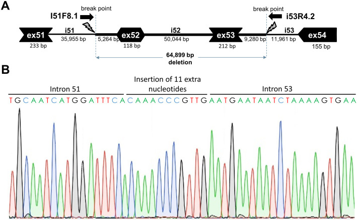
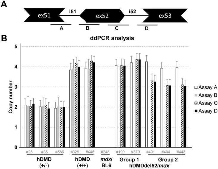
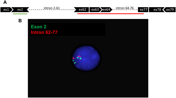
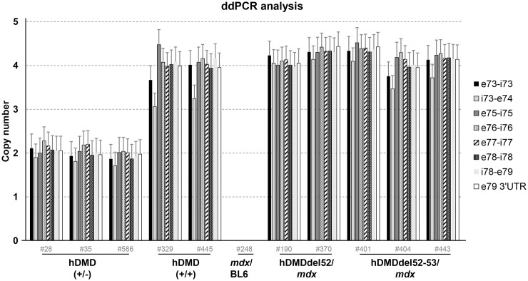
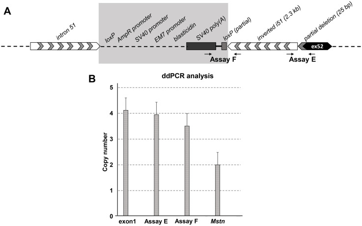
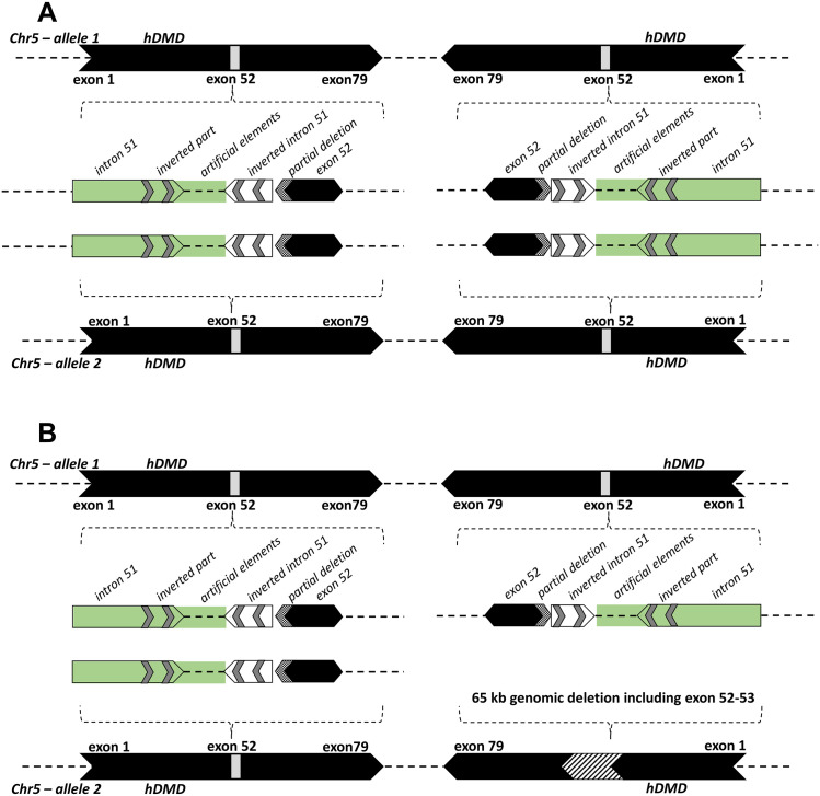
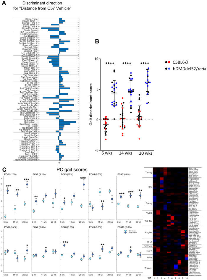
Duchenne muscular dystrophy (DMD) is a severe, progressive neuromuscular disorder caused by reading frame disrupting mutations in the DMD gene leading to absence of functional dystrophin. Antisense oligonucleotide (AON)-mediated exon skipping is a therapeutic approach aimed at restoring the reading frame at the pre-mRNA level, allowing the production of internally truncated partly functional dystrophin proteins. AONs work in a sequence specific manner, which warrants generating humanized mouse models for preclinical tests. To address this, we previously generated the hDMDdel52/mdx mouse model using transcription activator like effector nuclease (TALEN) technology. This model contains mutated murine and human DMD genes, and therefore lacks mouse and human dystrophin resulting in a dystrophic phenotype. It allows preclinical evaluation of AONs inducing the skipping of human DMD exons 51 and 53 and resulting in restoration of dystrophin synthesis. Here, we have further characterized this model genetically and functionally. We discovered that the hDMD and hDMDdel52 transgene is present twice per locus, in a tail-to-tail-orientation. Long-read sequencing revealed a partial deletion of exon 52 (first 25 bp), and a 2.3 kb inversion in intron 51 in both copies. These new findings on the genomic make-up of the hDMD and hDMDdel52 transgene do not affect exon 51 and/or 53 skipping, but do underline the need for extensive genetic analysis of mice generated with genome editing techniques to elucidate additional genetic changes that might have occurred. The hDMDdel52/mdx mice were also evaluated functionally using kinematic gait analysis. This revealed a clear and highly significant difference in overall gait between hDMDdel52/mdx mice and C57BL6/J controls. The motor deficit detected in the model confirms its suitability for preclinical testing of exon skipping AONs for human DMD at both the functional and molecular level.


Conflict of interest statement

AAR discloses being employed by LUMC which has patents on exon skipping technology, some of which has been licensed to BioMarin and subsequently sublicensed to Sarepta. As co-inventor of some of these patents AAR is entitled to a share of royalties. AAR further discloses being ad hoc consultant for PTC Therapeutics, Sarepta Therapeutics, CRISPR Therapeutics, Summit PLC, Alpha Anomeric, BioMarin Pharmaceuticals Inc., Eisai, Astra Zeneca, Santhera, Audentes, Global Guidepoint and GLG consultancy, Grunenthal, Wave and BioClinica, having been a member of the Duchenne Network Steering Committee (BioMarin) and being a member of the scientific advisory boards of ProQR, Sarepta, Silence and Philae Pharmaceuticals. Remuneration for these activities is paid to LUMC. LUMC also received speaker honoraria from PTC Therapeutics and BioMarin Pharmaceuticals and funding for contract research from Italpharmaco and Alpha Anomeric. RW, EK, CB, JvD and ND are (former) employees of BioMarin Nederland BV (formerly Prosensa Therapeutics BV) and performed the work with company budget in the form of salaries, equipment and facilities. JvD discloses being co-inventor on patents on exon skipping technology, some of which has been licensed to BioMarin and subsequently sublicensed to Sarepta. As co-inventor of some of these patents JvD is entitled to a share of royalties. JP, TB and TA are employees of Charles River Research Discovery Services in Finland and have no financial conflict of interest related to the submitted manuscript. Other authors have nothing to declare. The commercial affiliations of authors with Biomarin Pharmaceutical Ltd. and Charles River do not alter our adherence to PLOS ONE policies on sharing data and materials.

Figures

Fig 1
Fig 1. Unexpectedly high dystrophin levels detected in the heart of untreated and AON treated hDMDdel52/mdx mice.
A. Wes capillary immunoassay analysis revealed two groups of mice. Most mice had very low dystrophin levels (0.2–1.5%, mean: 0.7, standard deviation (SD): 0.2, group 1), while some treated and untreated mice had dystrophin levels in the range of 8.6–21.6% (mean: 13.4, SD: 3.0, group 2). B. Droplet digital qPCR revealed that exon skipping levels were comparable between the groups and therefore could not account for the differences observed in dystrophin protein levels. (Group 1: 0.1–4.8%, mean: 1.7, SD: 1.4 group 2: 0.2–4.2%, mean: 2.2, SD: 1.4). WT; wild type.
Fig 2
Fig 2. qPCR analysis of exon 51, 52 and 53 copy numbers.
A. Schematic representation of the probe locations. B. qPCR analysis of hDMD/mdx (+/- and +/+), mdx/BL6 and hDMDdel52 (group 1 and group 2) mice. To detect whether the hDMDdel52/mdx group 2 mice had an additional deletion of exon 51 or 53, qPCR was performed with primers targeting exon 51, 52 and 53 exon-intron boundaries. In both homozygous and heterozygous hDMD/mdx mice all exons could be detected, as expected with a 2-fold difference in copy number. For group 1 mice, exon 51 and exon 53 were detected, while exon 52 was absent. For the group 2 mice however, a reduced signal for exon 53 was detected.
Fig 3
Fig 3. Deletion breakpoint and sequence analysis.
A. Schematic depiction of the ~65 kb genomic deletion. B. Sanger sequence trace of the deletion breakpoint, including an insertion of 11 nucleotides at the break point.
Fig 4
Fig 4. ddPCR copy number analysis.
A. Schematic representation of the probe locations. B. ddPCR analysis of copy numbers of exons 51, 52 and 53 in hDMDdel52/mdx and hDMDdel52-53/mdx mice and controls, relative to Tfrc copy number. Most assays showed a double signal for both hDMD/mdx and hDMDdel52/mdx mice, while no signal was detected in mdx/BL6 mice. For the hDMDdel52/mdx and hDMDdel52-53/mdx mice, the probe on the intron 51- exon 52 boundary (assay B) did not give a signal. However, the probe on the exon 52 –intron 52 boundary did (assay C). As previously observed, assay C and assay D showed lower signals in hDMDdel52-53/mdx mice compared to hDMDdel52/mdx.
Fig 5
Fig 5. Detection of hDMD tandem duplication in hDMDdel52/mdx mouse by FISH analysis in interphase nuclei.
A. Schematic representation of FISH probes. B. Hybridization results under fluorescent microscopy. Two signals can be seen for the red probe which targets intron 62–77, while four signals are detected for the green probe targeting exon 2. Because metaphase spreads could not be detected, interphase nuclei were used for the analysis. hDMDdel52-53/mdx mice were not included in this analysis.
Fig 6
Fig 6. ddPCR analysis of copy numbers of exons 73 to 79 in hDMDdel52/mdx and hDMDdel52-53/mdx mice and controls, relative to Tfrc copy number.
Probes gave a double signal for hDMD, hDMDdel52/mdx and hDMDdel52-53/mdx mice, while no signal was detected in mdx/BL6 mice.
Fig 7
Fig 7. Genetic analysis of exon 52 deleted region.
A. Schematic representation of the exon 52 deletion region with flanking sites; the 25 bp partial deletion of exon 52 and 2.3 kb inversion of intron 51 were confirmed by sequencing. The map of the shaded area could not be confirmed by sequencing. B. ddPCR analysis of intron 51 inversion events in hDMD constructs. hDMDdel52/mdx (4 copies) samples were used in all assays. Data normalization was done relative to copy number of Mstn, which was set to 2 copies in hDMDdel52/mdx mice. Compared to the Mstn copy number, inversion specific primers in assay E showed a double signal while assay F gave lower signals in hDMDdel52/mdx mice, while they did not give any signals in hDMD/mdx mice as expected.
Fig 8
Fig 8. A schematic representation of hDMD gene constructs on mouse chromosome 5 of the hDMDdel52/mdx and hDMDdel52-53/mdx mice.
A. The figure represents the tail-to-tail duplication of the hDMD gene, and the genetic rearrangements which include a partial deletion of exon 52 and an inversion of part of intron 51 in the hDMDdel52/mdx mouse. The green area including intron 51 and artificial elements could not be confirmed by sequencing. B. Because the hDMD gene in the hDMDdel52-53/mdx mouse was not sequenced, the schematic representation is mostly hypothetical but ddPCR results revealed that a ~65 kb genomic deletion including exon 52–53 (Fig 3) occurred in one hDMD copy and the other three copies remained intact.
Fig 9
Fig 9. MotoRater analysis of hDMDdel52/mdx mice compared to C57BL6/J controls at the age of 6, 14 and 20 weeks.
A. Discriminant vector representing an overall kinematic fingerprint of the differences between hDMDdel52/mdx and C57BL6/J mice. B. Gait discriminant score reflecting the totality of gait parameters. Black dots represent male mice in each group, while blue and red dots represent females. Statistical significance was assessed per time point using an unpaired two-tailed t-test (****: P<0.0001). Statistical differences were more pronounced in female mice. C. Heatmap showing 10 PC clusters reflecting different gait features that differ between hDMDdel52/mdx and C57BL6/J mice. The right panel displays the gait scores for the different PCs. The % of the total variation of the data explained by each PC is indicated between brackets. Values are presented as mean ± standard error of mean (SEM). Significance per time point was assessed using an unpaired two-tailed t-test (*: P<0.05; **: P<0.01; ***: P<0.001). hDMDdel52-53/mdx mice were not included in MotoRater analysis.

References

    1. Moat SJ, Bradley DM, Salmon R, Clarke A, Hartley L. Newborn bloodspot screening for Duchenne muscular dystrophy: 21 years experience in Wales (UK). European journal of human genetics: EJHG. 2013;21(10):1049–53. Epub 2013/01/24. 10.1038/ejhg.2012.301 - DOI - PMC - PubMed
    1. Gao QQ, McNally EM. The Dystrophin Complex: Structure, Function, and Implications for Therapy. Compr Physiol. 2015;5(3):1223–39. Epub 2015/07/04. 10.1002/cphy.c140048 - DOI - PMC - PubMed
    1. Rader EP, Turk R, Willer T, Beltrán D, Inamori K-i, Peterson TA, et al. Role of dystroglycan in limiting contraction-induced injury to the sarcomeric cytoskeleton of mature skeletal muscle. 2016;113(39):10992–7. 10.1073/pnas.1605265113 %J Proceedings of the National Academy of Sciences. - DOI - PMC - PubMed
    1. Pane M, Scalise R, Berardinelli A, D’Angelo G, Ricotti V, Alfieri P, et al. Early neurodevelopmental assessment in Duchenne muscular dystrophy. Neuromuscular disorders: NMD. 2013;23(6):451–5. Epub 2013/03/29. 10.1016/j.nmd.2013.02.012 . - DOI - PubMed
    1. Shimizu-Motohashi Y, Miyatake S, Komaki H, Takeda S, Aoki Y. Recent advances in innovative therapeutic approaches for Duchenne muscular dystrophy: from discovery to clinical trials. American journal of translational research. 2016;8(6):2471–89. Epub 2016/07/12. - PMC - PubMed

LinkOut - more resources