

Alteration of Cortical and Subcortical Structures in Children With Profound Sensorineural Hearing Loss
- PMID: 33362488
- PMCID: PMC7756106
- DOI: 10.3389/fnhum.2020.565445
Alteration of Cortical and Subcortical Structures in Children With Profound Sensorineural Hearing Loss
Abstract
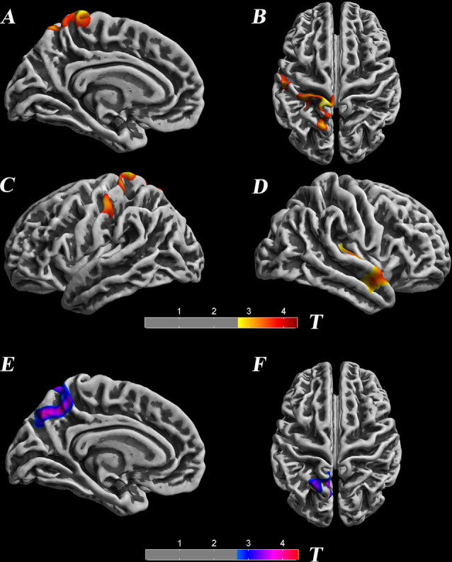
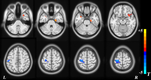
Profound sensorineural hearing loss (SNHL) is an auditory disability associated with auditory and cognitive dysfunction. Due to distinct pathogenesis, some associated structural and functional changes within the brain have been investigated in previous studies, but whole-brain structural alterations are incompletely understood. We extended the exploration of neuroanatomic differences in whole-brain structure in children with profound SNHL who are primarily users of Chinese sign language (CSL). We employed surface-based morphometry (SBM) and subcortical analyses. T1-weighted magnetic resonance images of 26 children with profound SNHL and 27 age- and sex-matched children with normal hearing were analyzed. Compared with the normal control (NC) group, children with profound SNHL showed diverse structural changes in surface-based and subcortical analyses, including decreased cortical thickness in the left postcentral gyrus, superior parietal lobule, paracentral lobule, precuneus, the right transverse temporal gyri, and the middle temporal gyrus; a noticeable increase in the Local Gyrification Index (LGI) in the left precuneus and superior parietal lobule; and diverse changes in gray-matter volume (GMV) in different brain regions. Surface-based vertex analyses revealed regional contractions in the right thalamus, putamen, pallidum, and the brainstem of children with profound SNHL when compared with those in the NC group. Volumetric analyses showed decreased volumes of the right thalamus and pallidum in children with profound SNHL. Our data suggest that children with profound SNHL are associated with diffuse cerebral dysfunction to cortical and subcortical nuclei, and revealed neuroplastic reorganization in the precuneus, superior parietal lobule, and temporal gyrus. Our study provides robust evidence for changes in connectivity and structure in the brain associated with hearing loss.
Keywords: children; multi-modal; sensorineural hearing loss; structural MRI; surface-based morphometry; surface-based vertex analysis.
Copyright © 2020 Qu, Tang, Pan, Zhao and Wang.
Conflict of interest statement
The authors declare that the research was conducted in the absence of any commercial or financial relationships that could be construed as a potential conflict of interest.
Figures

Similar articles
-
Cortical thickness analysis and optimized voxel-based morphometry in children and adolescents with prelingually profound sensorineural hearing loss.Brain Res. 2012 Jan 9;1430:35-42. doi: 10.1016/j.brainres.2011.09.057. Epub 2011 Oct 2. Brain Res. 2012. PMID: 22079323
-
The left lateral occipital cortex exhibits decreased thickness in children with sensorineural hearing loss.Int J Dev Neurosci. 2019 Aug;76:34-40. doi: 10.1016/j.ijdevneu.2019.05.009. Epub 2019 Jun 4. Int J Dev Neurosci. 2019. PMID: 31173823 Free PMC article.
-
White matter structural network alterations in congenital bilateral profound sensorineural hearing loss children: A graph theory analysis.Hear Res. 2022 Sep 1;422:108521. doi: 10.1016/j.heares.2022.108521. Epub 2022 May 16. Hear Res. 2022. PMID: 35660126
-
Gray Matter Structural Alterations in Social Anxiety Disorder: A Voxel-Based Meta-Analysis.Front Psychiatry. 2018 Sep 21;9:449. doi: 10.3389/fpsyt.2018.00449. eCollection 2018. Front Psychiatry. 2018. PMID: 30298028 Free PMC article. Review.
-
Cortical and Subcortical Gray Matter Volume in Youths With Conduct Problems: A Meta-analysis.JAMA Psychiatry. 2016 Jan;73(1):64-72. doi: 10.1001/jamapsychiatry.2015.2423. JAMA Psychiatry. 2016. PMID: 26650724 Review.
Cited by
-
Cerebral cortex functional reorganization in preschool children with congenital sensorineural hearing loss: a resting-state fMRI study.Front Neurol. 2024 Jun 25;15:1423956. doi: 10.3389/fneur.2024.1423956. eCollection 2024. Front Neurol. 2024. PMID: 38988601 Free PMC article.
-
Altered Functional Network in Infants With Profound Bilateral Congenital Sensorineural Hearing Loss: A Graph Theory Analysis.Front Neurosci. 2022 Jan 14;15:810833. doi: 10.3389/fnins.2021.810833. eCollection 2021. Front Neurosci. 2022. PMID: 35095404 Free PMC article.
-
Volumetric Analysis of Hearing-Related Structures of Brain in Children with GJB2-Related Congenital Deafness.Children (Basel). 2022 May 30;9(6):800. doi: 10.3390/children9060800. Children (Basel). 2022. PMID: 35740737 Free PMC article.
-
Accelerated Brain Atrophy, Microstructural Decline and Connectopathy in Age-Related Macular Degeneration.Biomedicines. 2024 Jan 10;12(1):147. doi: 10.3390/biomedicines12010147. Biomedicines. 2024. PMID: 38255252 Free PMC article.
-
Altered static and dynamic intrinsic brain activity in unilateral sudden sensorineural hearing loss.Front Neurosci. 2023 Aug 31;17:1257729. doi: 10.3389/fnins.2023.1257729. eCollection 2023. Front Neurosci. 2023. PMID: 37719156 Free PMC article.
References
LinkOut - more resources
Full Text Sources