

SDF-1α/MicroRNA-134 Axis Regulates Nonfunctioning Pituitary Neuroendocrine Tumor Growth via Targeting VEGFA
- PMID: 33362712
- PMCID: PMC7756115
- DOI: 10.3389/fendo.2020.566761
SDF-1α/MicroRNA-134 Axis Regulates Nonfunctioning Pituitary Neuroendocrine Tumor Growth via Targeting VEGFA
Abstract
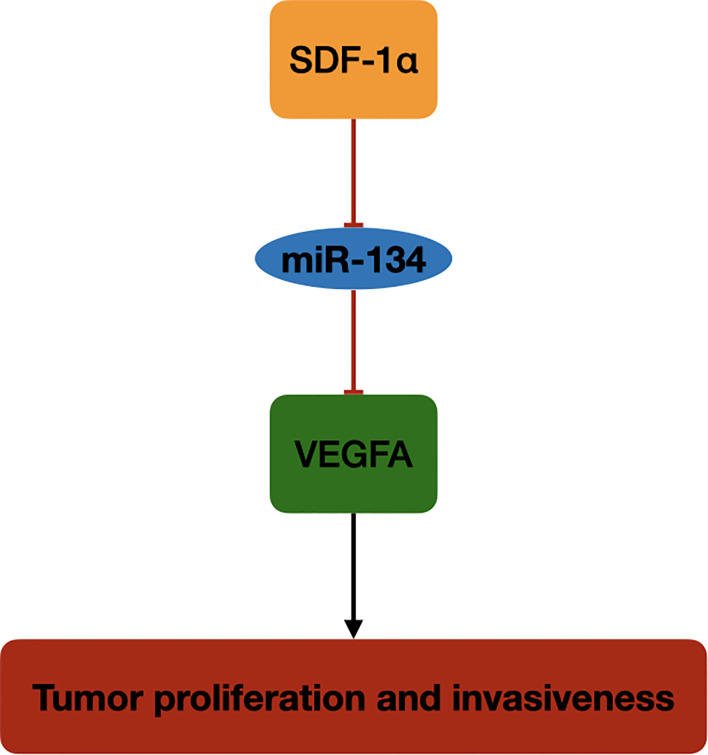
Background: Nonfunctioning pituitary neuroendocrine tumor (NF-PitNET) is difficult to resect. Except for surgery, there is no effective treatment for NF-PitNET. MicroRNA-134 (miR-134) has been reported to inhibit proliferation and invasion ability of tumor cells. Herein, the mechanism underlying the effect of miR-134 on alleviating NF-PitNET tumor cells growth is explored.
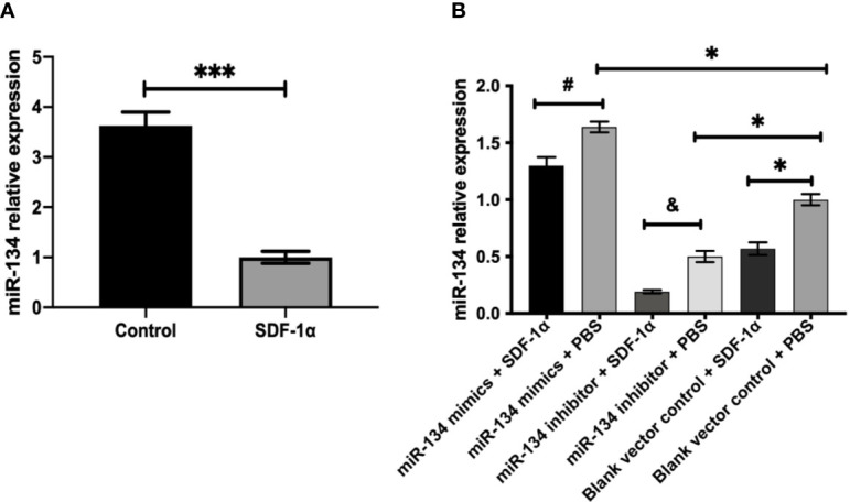
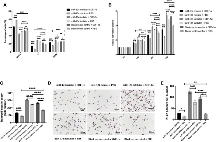
Methods: Mouse pituitary αT3-1 cells were transfected with miR-134 mimics and inhibitor, followed by treatment with stromal cell-derived factor-1α (SDF-1α) in vitro. MiR-134 expression level: we used quantitative real-time PCR (qRT-PCR) to detect the expression of miR-134. Cell behavior level: cell viability and invasion ability were assessed using a cell counting kit-8 (CCK8) assay and Transwell invasion assay respectively. Cytomolecular level: tumor cell proliferation was evaluated by Ki-67 staining; propidium iodide (PI) staining analyzed the effect of miR-134 on cell cycle arrest; western blot analysis and immunofluorescence staining evaluated tumor migration and invasive ability. Additionally, we collected 27 NF-PitNET tumor specimens and related clinical data. The specimens were subjected to qRT-PCR to obtain the relative miR-134 expression level of each specimen; linear regression analysis was used to analyze the miR-134 expression level in tumor specimens and the age of the NF-PitNET population, gender, tumor invasion, prognosis, and other indicators.
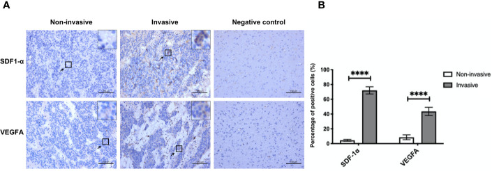
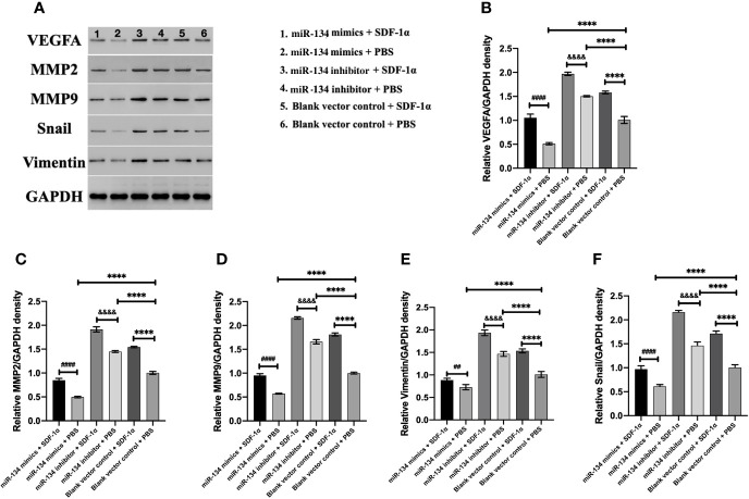
Results: In vitro experiment, miR-134 was observed to significantly inhibit αT3-1 cells proliferation characterized by inhibited cell viability and expressions of vascular endothelial growth factor A (VEGFA) and cell cycle transition from G1 to S phase (P < 0.01). VEGFA was verified as a target of miR-134. Additionally, miR-134-induced inhibition of αT3-1 cell proliferation and invasion was attenuated by SDF-1α and VEGFA overexpression (P < 0.01). In primary NF-PitNET tumor analysis, miR-134 expression level was negatively correlated with tumor invasion (P = 0.003).
Conclusion: The regulation of the SDF-1α/miR-134/VEGFA axis represents a novel mechanism in the pathogenesis of NF-PitNETs and may serve as a potential therapeutic target for the treatment of NF-PitNETs.
Keywords: SDF-1α (CXCL12); invasion; microRNA-134(miR-134); nonfunctioning pituitary neuroendocrine tumor (NF-PitNET); proliferation; vascular endothelial growth factor A (VEGFA).
Copyright © 2020 Wang, Fang, Zhou, Guo, Xu, Li, Zhang and Hong.
Conflict of interest statement
The authors declare that the research was conducted in the absence of any commercial or financial relationships that could be construed as a potential conflict of interest.
Figures
Similar articles
-
Differential expression levels of β-catenin are associated with invasive behavior of both functional and non-functional pituitary neuroendocrine tumor (PitNET).Mol Biol Rep. 2023 Aug;50(8):6425-6434. doi: 10.1007/s11033-023-08523-0. Epub 2023 Jun 16. Mol Biol Rep. 2023. PMID: 37326745
-
Lower 68 Ga-DOTATOC uptake in nonfunctioning pituitary neuroendocrine tumours compared to normal pituitary gland-A proof-of-concept study.Clin Endocrinol (Oxf). 2020 Mar;92(3):222-231. doi: 10.1111/cen.14144. Epub 2020 Jan 9. Clin Endocrinol (Oxf). 2020. PMID: 31868239
-
DNA hypomethylation-related expression of hsa-miR-184 contributes to invasive growth of gonadotroph neuroendocrine pituitary tumors.J Neuroendocrinol. 2025 Apr;37(4):e13492. doi: 10.1111/jne.13492. Epub 2025 Jan 23. J Neuroendocrinol. 2025. PMID: 39846216
-
Progress in the Pathogenesis, Diagnosis, and Treatment of TSH-Secreting Pituitary Neuroendocrine Tumor.Front Endocrinol (Lausanne). 2020 Nov 27;11:580264. doi: 10.3389/fendo.2020.580264. eCollection 2020. Front Endocrinol (Lausanne). 2020. PMID: 33329389 Free PMC article. Review.
-
Pituitary Tumors: Genetic and Molecular Factors Underlying Pathogenesis and Clinical Behavior.Neuroendocrinology. 2022;112(1):15-33. doi: 10.1159/000514862. Epub 2021 Feb 1. Neuroendocrinology. 2022. PMID: 33524974 Review.
Cited by
-
Regulatory mechanisms of microRNAs in endocrine disorders and their therapeutic potential.Front Genet. 2023 Feb 21;14:1137017. doi: 10.3389/fgene.2023.1137017. eCollection 2023. Front Genet. 2023. PMID: 36896239 Free PMC article. Review.
-
High-Grade Ectopic Pituitary Adenoma within the Cerebellopontine Angle: A Case Report.J Neurol Surg Rep. 2023 Apr 21;84(2):e51-e58. doi: 10.1055/a-2065-9809. eCollection 2023 Apr. J Neurol Surg Rep. 2023. PMID: 37090942 Free PMC article.
-
The Emerging Role of Non-Coding RNAs in Pituitary Gland Tumors and Meningioma.Cancers (Basel). 2021 Nov 28;13(23):5987. doi: 10.3390/cancers13235987. Cancers (Basel). 2021. PMID: 34885097 Free PMC article. Review.
-
A Retrospective Trail Investigating Temozolomide Neoadjuvant Chemotherapy Combined with Radiotherapy in Low-Grade Pituitary Tumors.J Oncol. 2022 Mar 25;2022:4618664. doi: 10.1155/2022/4618664. eCollection 2022. J Oncol. 2022. PMID: 35368902 Free PMC article.
-
Chemokine receptor 7 mediates miRNA-182 to regulate cerebral ischemia/reperfusion injury in rats.CNS Neurosci Ther. 2023 Feb;29(2):712-726. doi: 10.1111/cns.14056. Epub 2022 Dec 15. CNS Neurosci Ther. 2023. PMID: 36523152 Free PMC article.
References
Publication types
MeSH terms
Substances
LinkOut - more resources
Full Text Sources
Medical
Research Materials
Miscellaneous