Review
. 2020 Dec 11:11:608645.
doi: 10.3389/fimmu.2020.608645. eCollection 2020.

Importance of Type I and III Interferons at Respiratory and Intestinal Barrier Surfaces

Affiliations
Review

Importance of Type I and III Interferons at Respiratory and Intestinal Barrier Surfaces

Megan L Stanifer et al. Front Immunol. .

Abstract

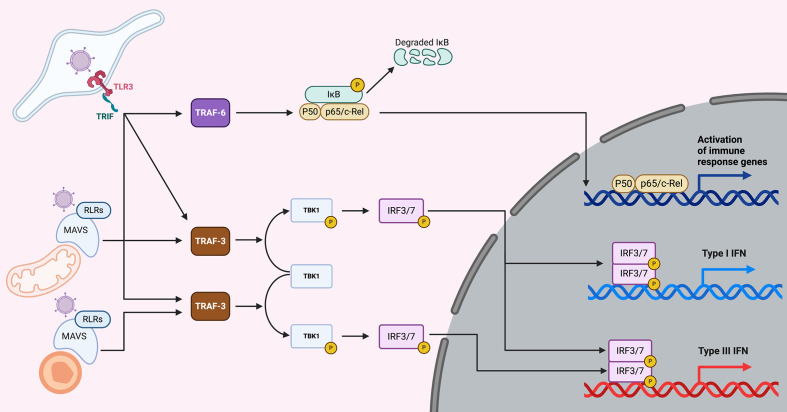
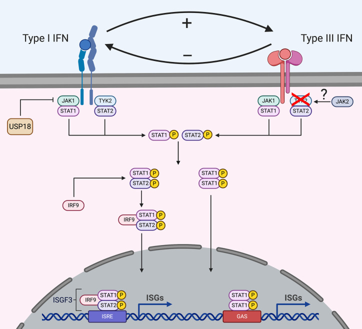
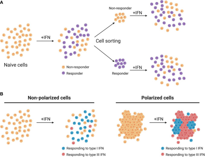
Interferons (IFNs) constitute the first line of defense against microbial infections particularly against viruses. They provide antiviral properties to cells by inducing the expression of hundreds of genes known as interferon-stimulated genes (ISGs). The two most important IFNs that can be produced by virtually all cells in the body during intrinsic innate immune response belong to two distinct families: the type I and type III IFNs. The type I IFN receptor is ubiquitously expressed whereas the type III IFN receptor's expression is limited to epithelial cells and a subset of immune cells. While originally considered to be redundant, type III IFNs have now been shown to play a unique role in protecting mucosal surfaces against pathogen challenges. The mucosal specific functions of type III IFN do not solely rely on the restricted epithelial expression of its receptor but also on the distinct means by which type III IFN mediates its anti-pathogen functions compared to the type I IFN. In this review we first provide a general overview on IFNs and present the similarities and differences in the signal transduction pathways leading to the expression of either type I or type III IFNs. By highlighting the current state-of-knowledge of the two archetypical mucosal surfaces (e.g. the respiratory and intestinal epitheliums), we present the differences in the signaling cascades used by type I and type III IFNs to uniquely induce the expression of ISGs. We then discuss in detail the role of each IFN in controlling pathogen infections in intestinal and respiratory epithelial cells. Finally, we provide our perspective on novel concepts in the field of IFN (stochasticity, response heterogeneity, cellular polarization/differentiation and tissue microenvironment) that we believe have implications in driving the differences between type I and III IFNs and could explain the preferences for type III IFNs at mucosal surfaces.

Keywords: epithelial cells; interferon lambda; interferons; intestinal epithelium; mucosal immunity; respiratory epithelia; type I interferon; type III interferon.


Conflict of interest statement

The authors declare that the research was conducted in the absence of any commercial or financial relationships that could be construed as a potential conflict of interest.

Figures

Figure 1
Figure 1
Overview of IFN production upon viral infection. Upon virus entry into cells, viruses are sensed by the TLRs (endosomes) or the RLRs (cytoplasm). TLR3 senses dsRNA (a main component of viruses or viral replication) and is located in endosomes. Upon sensing of dsRNA molecules, TLR3 and its adapter TRIF lead to the induction of both the NFкB and the IRF3/7 pathways, which lead to the induction of both pro-inflammatory cytokines and type I and III IFNs. Viral PAMPs located in the cytosol are recognized by RLRs and upon activation recruit the adapter protein MAVS. When MAVS is recruited to mitochondria, NFкB and IRF3/7 are activated leading to the induction of pro-inflammatory cytokines and type I and III IFNs. However, when MAVS is recruited to peroxisomes the signaling cascade leads to the induction of type III IFNs only.
Figure 2
Figure 2
Signal transduction downstream type I and type III IFN receptors. Upon binding to their receptors, IFNs induce the activation of the JAK/STAT signaling cascade. Both type I and III IFNs use JAK1 for their signaling, while type I IFNs also require TYK2 activation, type III IFNs signal independently of TYK2. Several studies suggest that type III IFNs use JAK2 for their signaling while type I interferons do not require JAK2. However, how JAK2 interacts with the receptor complex is currently unknown. Following JAK activation, STATs are recruited and activated which leads to their dimerization and binding to IRF9 forming the ISGF3 complex or homodimer complexes which translocates into the nucleus and drives ISG production. Some ISGs act as negative regulators and the ISG USP18 is known to regulate type I IFN signaling but not type III IFN signaling.
Figure 3
Figure 3
Heterogeneity of IFN production and response. (A) Homogeneous cell cultures treated with IFN (+IFN) respond in a heterogeneous manner. Upon sorting and restimulation with IFN, non-responding cells display a similar distribution of responding cells as the naive population. (B) In murine models, non-polarized cells respond mainly to type I IFNs while polarized cells respond mainly to type III IFNs.

References

    1. Isaacs A, Lindenmann J. Virus interference I. interferon. Proc R Soc Lond B Biol Sci (1957) 147:258–67. 10.1098/rspb.1957.0048 - DOI - PubMed
    1. Schoggins JW, Rice CM. Interferon-stimulated genes and their antiviral effector functions. Curr Opin Virol (2011) 1:519–25. 10.1016/j.coviro.2011.10.008 - DOI - PMC - PubMed
    1. Schoggins JW. Interferon-Stimulated Genes: What Do They All Do? Annu Rev Virol (2019) 6:567–84. 10.1146/annurev-virology-092818-015756 - DOI - PubMed
    1. Isaacs A, Lindenmann J. Virus interference. I. The interferon. By A. Isaacs and J. Lindenmann. J Interferon Res (1987) 7:429–38. 10.1089/jir.1987.7.429 - DOI - PubMed
    1. LaFleur DW, Nardelli B, Tsareva T, Mather D, Feng P, Semenuk M, et al. Interferon-kappa novel type I interferon expressed human keratinocytes. J Biol Chem (2001) 276:39765–71. 10.1074/jbc.M102502200 - DOI - PubMed

Publication types

LinkOut - more resources