Review
. 2020 Dec 9:10:602661.
doi: 10.3389/fonc.2020.602661. eCollection 2020.

Role of Autophagy in the Microenvironment of Oral Squamous Cell Carcinoma

Affiliations
Review

Role of Autophagy in the Microenvironment of Oral Squamous Cell Carcinoma

Daniel Peña-Oyarzún et al. Front Oncol. .

Abstract

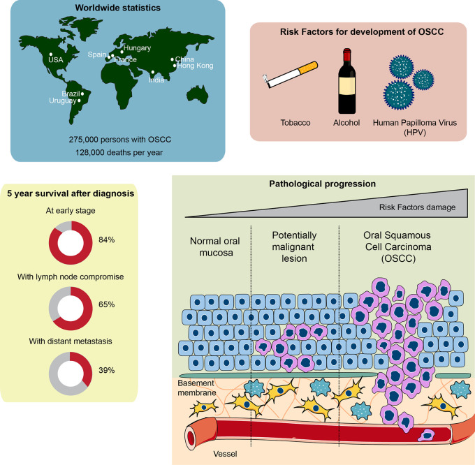
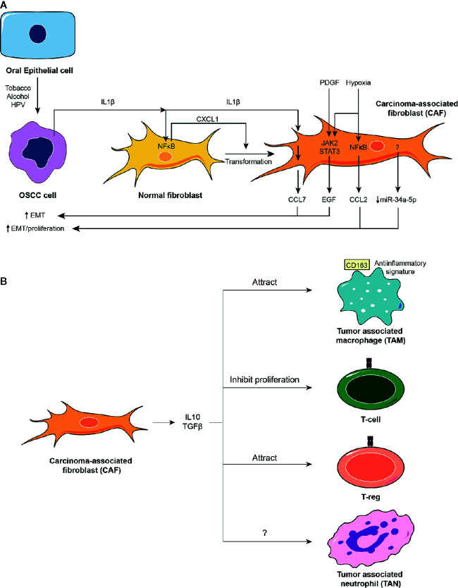
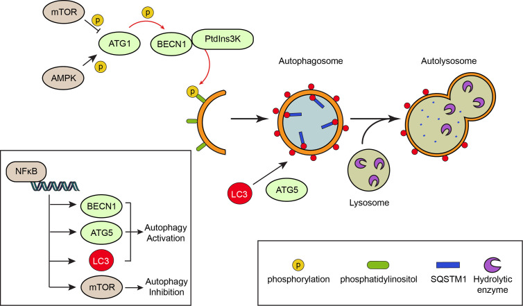
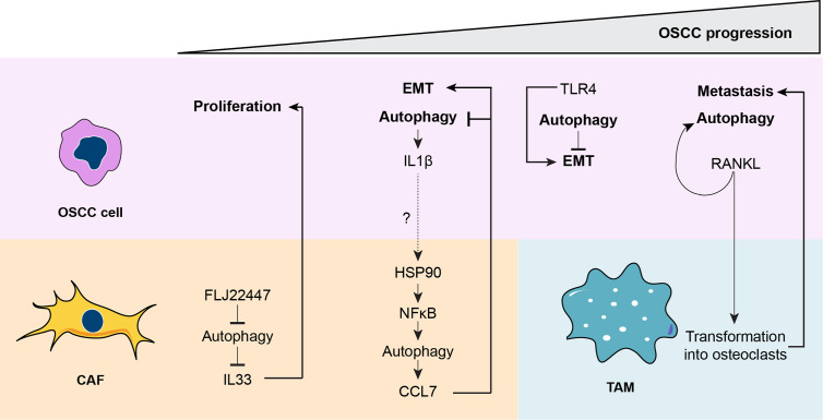
Oral squamous cell carcinoma, the most common type of oral cancer, affects more than 275,000 people per year worldwide. Oral squamous cell carcinoma is very aggressive, as most patients die after 3 to 5 years post-diagnosis. The initiation and progression of oral squamous cell carcinoma are multifactorial: smoking, alcohol consumption, and human papilloma virus infection are among the causes that promote its development. Although oral squamous cell carcinoma involves abnormal growth and migration of oral epithelial cells, other cell types such as fibroblasts and immune cells form the carcinoma niche. An underlying inflammatory state within the oral tissue promotes differential stress-related responses that favor oral squamous cell carcinoma. Autophagy is an intracellular degradation process that allows cancer cells to survive under stress conditions. Autophagy degrades cellular components by sequestering them in vesicles called autophagosomes, which ultimately fuse with lysosomes. Although several autophagy markers have been associated with oral squamous cell carcinoma, it remains unclear whether up- or down-regulation of autophagy favors its progression. Autophagy levels during oral squamous cell carcinoma are both timing- and cell-specific. Here we discuss how autophagy is required to establish a new cellular microenvironment in oral squamous cell carcinoma and how autophagy drives the phenotypic change of oral squamous cell carcinoma cells by promoting crosstalk between carcinoma cells, fibroblasts, and immune cells.

Keywords: autophagy; cancer; carcinoma-associated fibroblast; oral squamous cell carcinoma; tumor microenvironment.


Conflict of interest statement

The authors declare that the research was conducted in the absence of any commercial or financial relationships that could be construed as a potential conflict of interest.

Figures

Figure 1
Figure 1
Global statistics, survival rate, and pathophysiological features of oral squamous cell carcinoma (OSCC). Top left: countries with higher cases of OSCC diagnosed around the world. Top right: the main risk factors involved in OSCC development and progression. Bottom left: chances of survival 5 years after being diagnosed with OSCC. Note that early diagnosis of OSCC is crucial to ensure over 80% survival chance after 5 years. The general statistics show a 50% survival rate after 5 years, given that OSCC is usually diagnosed late. Bottom right: development of OSCC from a normal oral epithelium. The normal epithelium, composed of epithelial cells known as keratinocytes, is located over a basement membrane that separates the epithelium from the connective tissue composed of fibroblasts, immune cells and vessels. Exposure to carcinogens derived from the risk factors of the top right panel generate a potentially malignant lesion, characterized by an altered cellular morphology that starts affecting the inner layers of the epithelium close to the basement membrane, progressing toward the outer layers of the epithelium. Continuous exposure to carcinogens leads to OSCC development, a phenomenon that alters all the epithelial cell layers both genetically and morphologically. Interplay between connective tissue cells and OSCC cells is also observed, which assists OSCC growth and metastasis.
Figure 2
Figure 2
The oral squamous cell carcinoma (OSCC) tumor microenvironment. (A) The OSCC tumor microenvironment is mainly composed of cancer-associated fibroblasts (CAFs). CAFs derive from normal fibroblasts after autocrine stimulation of chemokine (C-X-C motif) ligand (CXCL1) chemokine through a nuclear factor κB (NFκB)-dependent mechanism. NFκB is activated in fibroblasts by the action of interleukin-1β (IL1β) released from OSCC cells. Conditions surrounding the tumoral tissue, such as increased levels of platelet-derived growth factor (PDGF) and IL1β or hypoxia activate Janus kinase (JAK)/STAT and NFκB pathways in CAFs, which induce the release of chemokines (CCL2 and CCL7) and epithelial growth factor (EGF), or inhibit the release of selective miRs such as miR-34a-5p. All these mediators augment proliferation and EMT of OSCC cells. (B) CAFs release well known anti-inflammatory molecules such as IL10 and TGFβ that attract anti-inflammatory macrophages and regulatory T-cells, T-regs, and inhibit proliferation of T-cells.
Figure 3
Figure 3
The mechanism of autophagy. Autophagy is a degradation process that involves the formation of double membrane vesicles called autophagosomes. Canonical signaling pathways, such as activation of the nutrient deprivation sensor AMP-activated protein kinase (AMPK) or inhibition of the nutrient-full sensor mechanistic target of rapamycin (mTOR) activate autophagy-related protein 1 (ATG1) kinase by phosphorylation. This in turns phosphorylates the Beclin 1 (BECN1) protein, allowing formation of a PtdIns3K complex that phosphorylates the phosphatidylinositol of intracellular membranes. Formation of the autophagosomes implicates the elongation of the membranes and their decoration with LC3 molecules, a process that is assisted by the ATG5 protein. The material targeted for degradation (i.e. proteins and organelles) arrives to the elongating membrane of the autophagosome through “receptors”, like SQSTM1/p62, which binds polyubiquitinated proteins. Once the autophagosome is formed, it fuses with a lysosome that contains hydrolytic enzymes, leading to the degradation of the material enclosed within the autophagosome. The NFκB pathway is involved in transcriptional up-regulation of autophagy proteins such as BECN1, LC3 and ATG5, thereby promoting autophagy.
Figure 4
Figure 4
A unified model for the role of autophagy during oral squamous cell carcinoma (OSCC) progression. During the early stages of OSCC, the long non-coding RNA FLJ22447 inhibits autophagy in cancer-associated fibroblasts (CAFs), impairing the autophagic degradation of IL33. Increased levels of IL33 are released from CAFs, inducing proliferation of the OSCC cells. Then, OSCC cells increase autophagy that releases interleukin-1β (IL1β), which then increases autophagy in CAFs through an nuclear factor κB ((NFκB)-dependent mechanism. The IL1β released from the OSCC cells may promote activation of heat shock protein 90 (HSP90) in the CAFs to increase NFκB activity, but this needs to be demonstrated (dashed line). However, it is known that NFκB-dependent autophagy in CAFs induces release of chemokine (C-C motif) ligand 7 (CCL7), which acts on OSCC cells, promoting epithelial-mesenchymal transition (EMT) and inhibiting autophagy. During later phases of OSCC, autophagy reduces Toll-like receptor 4 (TLR4)-dependent EMT. Therefore, inhibition of autophagy by CCL7 may promote TLR4-depedent EMT. Finally, during the advanced stages of OSCC, receptor activator of nuclear factor κB ligand (RANKL), which induces autophagy in OSCC cells, is released from OSCC cells transforming tumor-associated macrophages, TAMs, into osteoclasts, ultimately inducing metastasis.

Similar articles

Cited by

References

    1. Roma-Rodrigues C, Mendes R, Baptista PV, Fernandes AR. Targeting Tumor Microenvironment for Cancer Therapy. Int J Mol Sci (2019) 20(4):840. 10.3390/ijms20040840 - DOI - PMC - PubMed
    1. Ribeiro Franco PI, Rodrigues AP, de Menezes LB, Pacheco Miguel M. Tumor microenvironment components: Allies of cancer progression. Pathol Res Pract (2020) 216(1):152729. 10.1016/j.prp.2019.152729 - DOI - PubMed
    1. Rivera C. Essentials of oral cancer. Int J Clin Exp Pathol (2015) 8(9):11884–94. 10.5281/zenodo.192487 - DOI - PMC - PubMed
    1. Singh SS, Vats S, Chia AY, Tan TZ, Deng S, Ong MS, et al. Dual role of autophagy in hallmarks of cancer. Oncogene (2018) 37(9):1142–58. 10.1038/s41388-017-0046-6 - DOI - PubMed
    1. Patil S, Rao RS, Raj AT. Dual Role of Autophagy in Oral Cancer. J Int Oral Health (2015) 7(6):i–ii. - PMC - PubMed

LinkOut - more resources