Review
. 2020 Dec 10:8:599454.
doi: 10.3389/fbioe.2020.599454. eCollection 2020.

Disentangling Biomolecular Corona Interactions With Cell Receptors and Implications for Targeting of Nanomedicines

Affiliations
Review

Disentangling Biomolecular Corona Interactions With Cell Receptors and Implications for Targeting of Nanomedicines

Aldy Aliyandi et al. Front Bioeng Biotechnol. .

Abstract

Nanoparticles are promising tools for nanomedicine in a wide array of therapeutic and diagnostic applications. Yet, despite the advances in the biomedical applications of nanomaterials, relatively few nanomedicines made it to the clinics. The formation of the biomolecular corona on the surface of nanoparticles has been known as one of the challenges toward successful targeting of nanomedicines. This adsorbed protein layer can mask targeting moieties and creates a new biological identity that critically affects the subsequent biological interactions of nanomedicines with cells. Extensive studies have been directed toward understanding the characteristics of this layer of biomolecules and its implications for nanomedicine outcomes at cell and organism levels, yet several aspects are still poorly understood. One aspect that still requires further insights is how the biomolecular corona interacts with and is "read" by the cellular machinery. Within this context, this review is focused on the current understanding of the interactions of the biomolecular corona with cell receptors. First, we address the importance and the role of receptors in the uptake of nanoparticles. Second, we discuss the recent advances and techniques in characterizing and identifying biomolecular corona-receptor interactions. Additionally, we present how we can exploit the knowledge of corona-cell receptor interactions to discover novel receptors for targeting of nanocarriers. Finally, we conclude this review with an outlook on possible future perspectives in the field. A better understanding of the first interactions of nanomaterials with cells, and -in particular -the receptors interacting with the biomolecular corona and involved in nanoparticle uptake, will help for the successful design of nanomedicines for targeted delivery.

Keywords: biomolecular corona; cell receptors; corona-receptor interactions; nanoparticles; targeting; uptake.


Conflict of interest statement

The authors declare that the research was conducted in the absence of any commercial or financial relationships that could be construed as a potential conflict of interest.

Figures
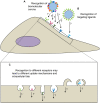
FIGURE 1
FIGURE 1
Interactions of nanoparticles with cell receptors. Recognition can be achieved via the biomolecular corona (A) and/or targeting ligands (B) (Allen, 2002; Lara et al., 2017; Francia et al., 2019; Villaverde and Baeza, 2019). Depending on which receptors nanoparticles interact with, uptake mechanisms may differ (here illustrated by different shapes of the membrane invaginations and membrane protrusions), which could affect the intracellular fate of nanoparticles (C).
FIGURE 2
FIGURE 2
Methods to elucidate nanoparticle protein corona-receptor interactions. First, corona proteins involved in nanoparticle uptake can be identified by associating the corona composition and nanoparticle uptake efficiency, for instance by using corona fingerprinting or correlation analysis (A) (Walkey et al., 2014; Liu et al., 2015; Ritz et al., 2015). The distribution of corona proteins and how their epitopes are presented can be determined by immunomapping-based techniques (B) (Kelly et al., 2015). Finally, receptors involved in the uptake of nanoparticle-corona complexes can be deduced from the corona composition or can be directly identified using pull-down or live-cell co-internalized receptor isolation approaches (C) (Bewersdorff et al., 2017; Ito et al., 2020; Aliyandi et al., unpublished).
FIGURE 3
FIGURE 3
Schematic illustration of the identification of nanoparticle corona proteins that are associated with cellular uptake. (1) A library of different nanoparticle formulations is incubated with a protein mixture in order to form a protein “fingerprint.” (2) The adsorbed proteins are isolated from the surface of the nanoparticles and (3) characterized by mass spectrometry. The serum protein fingerprint is a quantitative representation of each nanoparticle formulation. (4) Nanoparticles are incubated with cells. (5) Net cell association is determined. (6) A function “Y = f(X)” can be used to relate the corona composition to cell association, and f(X) can be used to predict the cell association of a certain nanoparticle formulation from its protein fingerprint. Adapted with permission from Walkey et al. (2014)–Copyright (2014) American Chemical Society.
FIGURE 4
FIGURE 4
Mapping of receptor binding motifs on the biomolecular corona and identification of receptors mediating uptake by cells. Schematic illustration of epitope mapping of ApoB-100 on the biomolecular corona of SiO2 nanoparticles by 5 nm immunogold nanoparticles conjugated with antibody anti-ApoB100 (A). Electron micrographs of ApoB-100 epitopes on the hard corona of SiO2 nanoparticles formed in 50% human serum and subsequently exposed to 50% delipidized serum for 4 h (B). Scheme of the low-density lipoprotein receptor LDLR fused with a fluorescently labeled HaloTag protein at its N-terminus (C). Uptake of nanoparticle-corona complexes in 50% delipidized serum in LDLR-transfected cells and control cells (Empty) (D). Uptake of nanoparticles-corona complexes can be competed with LDL in LDLR-transfected cells (E). All panels in this Figure are adapted with permission from Lara et al. (2017)–Copyright (2017) American Chemical Society.
FIGURE 5
FIGURE 5
Schematic illustration of the isolation of biotinylated cell surface proteins and the subsequent pull-down of proteins interacting with nanoparticle-corona complexes. First, cell surface proteins are labeled with biotin at 4°C. Next, the labeled surface proteins are purified and incubated with nanoparticle-corona complexes. Finally, cell surface proteins that are pulled-down by the nanoparticles are identified by mass spectrometry. Bewersdorff et al. (2017). Adapted from Aliyandi et al. (unpublished).
FIGURE 6
FIGURE 6
Schematic illustration of the method to isolate receptors internalized upon exposure to (biomolecular corona-coated) nanoparticles. First, cell surface proteins are labeled with biotin at 4°C. Next, the cells are incubated with nanoparticle-corona complexes at 37°C for a certain period of time to allow nanoparticle internalization. Then, the cell surface biotin is removed, and finally, labeled proteins that are internalized by the cell upon exposure to the nanoparticles are isolated and purified, and later identified with mass spectrometry. The same method is used to identify internalized receptors in control cells not exposed to the nanoparticles, in order to select only those internalized upon exposure to the nanoparticles. Adapted from Aliyandi et al. (unpublished).

References

    1. Akinc A., Querbes W., De S., Qin J., Frank-Kamenetsky M., Jayaprakash K. N., et al. (2010). Targeted delivery of RNAi therapeutics with endogenous and exogenous ligand-based mechanisms. Mol. Ther. 18 1357–1364. 10.1038/mt.2010.85 - DOI - PMC - PubMed
    1. Albanese A., Tang P. S., Chan W. C. W. (2012). The effect of nanoparticle size, shape, and surface chemistry on biological systems. Annu. Rev. Biomed. Eng. 14 1–16. 10.1146/annurev-bioeng-071811-150124 - DOI - PubMed
    1. Alberg I., Kramer S., Schinnerer M., Hu Q., Seidl C., Leps C., et al. (2020). Polymeric nanoparticles with neglectable protein corona. Small 16 1907574. 10.1002/smll.201907574 - DOI - PubMed
    1. Alexis F., Pridgen E., Molnar L. K., Farokhzad O. C. (2008). Factors affecting the clearance and biodistribution of polymeric nanoparticles. Mol. Pharm. 5 505–515. 10.1021/mp800051m - DOI - PMC - PubMed
    1. Allen T. M. (2002). Ligand-targeted therapeutics in anticancer therapy. Nat. Rev. Cancer 2 750–763. 10.1038/nrc903 - DOI - PubMed

LinkOut - more resources