. 2020 Dec 8:11:580547.
doi: 10.3389/fimmu.2020.580547. eCollection 2020.

Single-Stranded Oligonucleotide-Mediated Inhibition of Respiratory Syncytial Virus Infection

Affiliations

Single-Stranded Oligonucleotide-Mediated Inhibition of Respiratory Syncytial Virus Infection

Sandra Axberg Pålsson et al. Front Immunol. .

Abstract

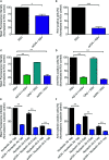
Respiratory syncytial virus (RSV) is the leading cause of acute lower respiratory tract infections in young children. Currently, there is no RSV vaccine or universally accessible antiviral treatment available. Addressing the urgent need for new antiviral agents, we have investigated the capacity of a non-coding single-stranded oligonucleotide (ssON) to inhibit RSV infection. By utilizing a GFP-expressing RSV, we demonstrate that the ssON significantly reduced the proportion of RSV infected A549 cells (lung epithelial cells). Furthermore, we show that ssON's antiviral activity was length dependent and that both RNA and DNA of this class of oligonucleotides have antiviral activity. We reveal that ssON inhibited RSV infection by competing with the virus for binding to the cellular receptor nucleolin in vitro. Additionally, using a recombinant RSV that expresses luciferase we show that ssON effectively blocked RSV infection in mice. Treatment with ssON in vivo resulted in the upregulation of RSV-induced interferon stimulated genes (ISGs) such as Stat1, Stat2, Cxcl10, and Ccl2. This study highlights the possibility of using oligonucleotides as therapeutic agents against RSV infection. We demonstrate that the mechanism of action of ssON is the inhibition of viral entry in vitro, likely through the binding of the receptor, nucleolin and that ssON treatment against RSV infection in vivo additionally results in the upregulation of ISGs.

Keywords: ISGs; antiviral; nucleolin; oligonucleotides; respiratory syncytial virus (RSV); single-stranded oligonucleotides; ssON.


Conflict of interest statement

AD and A-LS are shareholders of TIRmed Pharma in possession of intellectual properties related to ssON. A-LS is CEO of TIRmed Pharma. The remaining authors declare that the research was conducted in the absence of any commercial or financial relationships that could be construed as a potential conflict of interest.

Figures

Figure 1
Figure 1
Anti-respiratory syncytial virus (RSV) activity of ssON in vitro. A549 cells were infected with a RSV expressing GFP (RSV-GFP), at the indicated multiplicities of infection (MOI) for 2 h. Cells were further incubated for indicated time points prior to staining with LIVE/DEAD® Fixable near-IR Dead Cell Stain Kit (A). Cells were either pre-treated with a 35 bases long PS-stabilized ssON (ssON) for 2h or ssON (1 µM) was added simultaneously with RSV (MOI 1). The proportion of GFP positive live cells was determined 72 h post infection using flow cytometry (B). ssON was added to cells 2h before or concomitantly with RSV infection (MOI 1, 0.1, 0.05 or 0.01). The proportion of GFP positive live cells were measured 72 h and 96 h post infection using flow cytometry (C). The viability of RSV and ssON treated cells was measured 72 h and 96 h post infection using flow cytometry (D). Cells were treated with a 35 bases long un-stabilized ssON PO (ssON 35 PO) (1 µM) or PS-ssONs (1 µM) of different lengths as indicated by a number (ssON 30 PS etc). Alternatively, cells were treated with a 35 bases long single-stranded RNA with stabilizing modifications (ssON 2´OMe PS) 2 h before RSV infection at MOI 0.05. The proportion of live infected cells was measured 72 h post infection by flow cytometry. A549 cells were treated with (E) ssON (F), TMC353121 or (G) Presatovir (GS-5806) at indicated concentrations for 2h prior to RSV infection (MOI 0.05). The proportion to live infected cells was measured 72h post infection by flow cytometry and the IC50s are depicted in the graphs. Results are presented as mean ±SEM from three independent experiments in duplicates for each time point for all graphs. The statistical significance was measured using the non-parametric Mann-Whitney test for A, B, and C; all treatments were compared to the RSV infected untreated control for statistics. For G, H, and I the statistical significance was measured using the Kruskal-Wallis one-way ANOVA test with Dunns multiple comparison between each treatment conditions and the untreated RSV infected cells (control). P-value: P > 0.05; *P ≤ 0.05; **P ≤ 0.01. Lack of significance is not depicted in the figure.
Figure 2
Figure 2
ssON binds to cell surface expressed nucleolin. A549 cells were treated with ssON (1 µM), AS1411 (25 µM), or CRO (25 µM) for 30 min prior to incubation with respiratory syncytial virus (RSV) (MOI 2) for 1h at 4°C followed by detection with an anti-RSV F protein mAb combined with a APC-conjugated rat anti-mouse IgG1 secondary antibody. Cell surface bound RSV was subsequently detected by flow cytometry (A). Mean fluorescence intensity (MFI) of bound RSV in the presence or absence of ssON (B). Proportion of RSV positive cells in the presence or absence of ssON (C). MFI of cell surface bound RSV in the presence or absence of AS1411, CRO, or ssON (D). Proportion of RSV positive cells in the presence or absence of AS1411, CRO, or ssON. The data was normalized to the untreated RSV control. Alternatively, cells were treated with ssON (1 µM) for 30 min prior to incubation with an anti-nucleolin antibody at the indicated concentrations for 1 h at 4°C followed by staining with an Alexa 488-conjugated donkey anti-rabbit IgG secondary antibody (E). MFI of cell surface expressed nucleolin and (F) the proportion of cells labeled with an anti-nucleolin mAb was measured using flow cytometry. The data was normalized to the nucleolin 1 µg control. Results are presented as mean ±SEM from three to four independent experiments in duplicates for all graphs. The statistical significance for all graphs was measured using the non-parametric Mann-Whitney test and all treatments were compared to the RSV control. P-value: P > 0.05; *P ≤ 0.05; **P ≤ 0.01; ***P ≤ 0.001. Lack of significance is not depicted in the figure.
Figure 3
Figure 3
Treatment with ssON inhibits respiratory syncytial virus (RSV) infection in vivo in mice. Mice were infected intranasally with RSV-luciferase (2.106 pfu/ml) according to the schedule depicted in (A). Mice were given PBS or ssON (12.5 µg or 6.25 µg) intranasally 6 h before infection. A second treatment of ssON was administrated intranasally on day 2. The bioluminescence was measured using a in vivo system imaging (IVIS) after intranasal administration of D-luciferin, and capturing photon emission on day 0, 2, 3, and 4 post infection. Representative pictures of bioluminescence measured on the ventral view of mice after 4 days of infection with (B) RSV (C), RSV + ssON (6.25 µg), and (D) RSV + ssON (12.5 µg). The scale indicates the radiance in photons per second per square centimeter per steradian (p/s/cm2/sr). The “Living Image” software was used to quantify the luciferase activity (expressed as photon per seconds) on day 4 in (E) nose and (F) lungs ()* = outlier which was excluded from the statistical analysis. (G). The luciferase activity of the entire mouse was measured at indicated time points. Two independent experiments were performed and data show mean ±SEM with n=12 mice for each time point for all graphs. The statistical significance for all graphs was measured using an unpaired t-test with Welch’s correction comparing each treatment to the RSV control. P-value: P > 0.05; *P ≤ 0.05; **P ≤ 0.01; ****P ≤ 0.0001. Lack of significance is not depicted in the figure.
Figure 4
Figure 4
ssON treatment in vivo induces a differential immune profile in the lungs of respiratory syncytial virus (RSV) infected mice. RNA was extracted from the right lung lobes of mice treated with PBS, infected with RSV or from RSV infected mice treated with ssON (6.5 µg) 4 days post infection and analysed using Nanostring technology. Data consist of 9–10 mice per treatment from two independent experiments. DESeq2 analysis was used to normalize the immune gene expression to the internal reference genes. Genes with an adjusted p-value below 0.05 are fully colored (Significantly upregulated genes = green, significantly downregulated genes = red, rest = black) (A). MA-plots showing the expression profile in log2 fold change (L2FC) of immune genes in RSV infected mice compared to PBS treated mice. The top 10 most upregulated genes by L2FC are displayed (B). MA-plots showing the expression profile in L2FC of immune genes in ssON treated RSV infected mice compared to RSV infected mice. The top 10 most upregulated genes by L2FC are displayed.
Figure 5
Figure 5
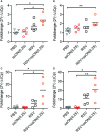
Treatment with ssON against respiratory syncytial virus (RSV) infection in vivo in mice upregulates the expression of interferon stimulated genes (ISGs). The expression of selected ISGs in the murine lungs was validated using RT-qPCR (A). Stat1 (B) Stat2 (C) Ccl2 (D) Cxcl10. Plots show mean from individual animals. Statistical significance was measured using the Kruskal-Wallis one-way ANOVA test with Dunns multiple comparison between each treatment conditions. P-value: P > 0.05; * P ≤ 0.05; ** P ≤ 0.01. Lack of significance is not depicted in the figures.

Similar articles

Cited by

References

    1. Collins PL, Fearns R, Graham BS. “Respiratory Syncytial Virus: Virology, Reverse Genetics, and Pathogenesis of Disease”. In: Anderson LJ, Graham BS, editors. Challenges and Opportunities for Respiratory Syncytial Virus Vaccines. Berlin, Heidelberg: Springer Berlin Heidelberg; (2013). p. 3–38. 10.1007/978-3-642-38919-1_1 - DOI - PMC - PubMed
    1. Karppinen S, Toivonen L, Schuez-Havupalo L, Waris M, Peltola V. Interference between respiratory syncytial virus and rhinovirus in respiratory tract infections in children. Clin Microbiol Infect (2016) 22(Supplement_7):208.e1–6. 10.1016/j.cmi.2015.10.002 - DOI - PubMed
    1. Shi T, Ooi Y, Zaw EM, Utjesanovic N, Campbell H, Cunningham S, et al. Association Between Respiratory Syncytial Virus-Associated Acute Lower Respiratory Infection in Early Life and Recurrent Wheeze and Asthma in Later Childhood. J Infect Dis (2019) 2(Issue Supplement 7):jiz311. 10.1093/infdis/jiz311 - DOI - PubMed
    1. Heylen E, Neyts J, Jochmans D. Drug candidates and model systems in respiratory syncytial virus antiviral drug discovery. Biochem Pharmacol (2017) 127:1–12. 10.1016/j.bcp.2016.09.014 - DOI - PubMed
    1. Shadman KA, Wald ER. A review of palivizumab and emerging therapies for respiratory syncytial virus. Expert Opin Biol Ther (2011) 11:1455–67. 10.1517/14712598.2011.608062 - DOI - PubMed

Publication types

MeSH terms