. 2020 Dec 8:11:586856.
doi: 10.3389/fimmu.2020.586856. eCollection 2020.

Methods to Detect MHC-Specific IgE in Mice and Men

Affiliations

Methods to Detect MHC-Specific IgE in Mice and Men

Anna Marianne Weijler et al. Front Immunol. .

Abstract

Humoral immunity is a major barrier limiting long-term outcome after organ transplantation. Especially, the production of antibodies directed against donor HLA/MHC antigens (i.e. donor-specific antibodies (DSA)) leading to antibody-mediated rejection (ABMR) is considered to be a major factor negatively affecting allograft survival. DSAs of the IgG isotype are routinely measured in transplant patients. However, not all patients diagnosed with IgG-DSA develop ABMR events. Therefore, research in better understanding the mechanisms of ABMR is of great importance. We recently demonstrated the production of MHC-specific IgE upon allograft rejection in mice and in transplant patients. IgE is classically connected with allergy and is known to be important for the humoral defense against helminths and worms. However, its role in autoimmune diseases and cancer has been reported recently as well. The concentration of IgE in blood is extremely low compared to other antibody isotypes. Therefore, detection of MHC-specific IgE from serum requires methods of high sensitivity. Since MHC-specific IgG-typically present at much higher serum levels-develops as well, high specificity is also required of IgE detection methods. In the murine model we developed an enzyme linked immunosorbent assay (ELISA) using MHC monomers for measurement of MHC-specific IgE, allowing us to distinguish between specificities of antibodies against different class I and class II antigens. For measurement of functional activity of MHC-specific IgE in vitro, a release assay using a rat basophil cell line (RBL-2H3) was established. For functional analysis of MHC-specific IgE in vivo, a cutaneous hypersensitivity reaction assay was adapted for this purpose using MHC monomers. Humanized RBL-2H3 cells transfected with cDNA coding for the human-high affinity IgE receptor were used for functionality measurement of donor-specific IgE in sensitized transplant patients. For detection of HLA-specific IgE, a bead assay was adapted, using beads expressing single HLA antigens. The aim of this publication is to demonstrate currently established methods for the detection and characterization of MHC-specific IgE in the murine and human setting.

Keywords: HLA; IgE; MHC; antibody-mediated rejection; donor specific antibodies; transplantation.


Conflict of interest statement

The authors declare that the research was conducted in the absence of any commercial or financial relationships that could be construed as a potential conflict of interest.

Figures
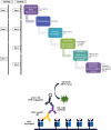
Figure 1
Figure 1
Step by step procedure and schematic representation of the MHC-specific ELISA. Murine MHC monomers are incubated with murine serum to be analyzed for the presence of MHC-specific IgE. MHC-specific antibodies bind to their respective epitope on the monomers and are then detected using a primary antibody. The timeline of the ELISA procedure varies with the incubation time and temperature of the primary antibody. When incubated for 2 h at RT the ELISA takes three days (timeline 1), when incubated at 4°C overnight, the ELISA takes four days (timeline 2). The secondary or detection antibody is conjugated with HRP. Due to the presence of hydrogen peroxide and HRP, ABTS is oxidized to a radical cation. The resulting blue-green reaction product of ABTS can be measured at 405 nm.
Figure 2
Figure 2
Step by step procedure and schematic representation of the rat basophil leukemia cell degranulation assay. Cells from the rat basophilic leukemia strain (RBL-2H3) are incubated with murine serum that shall be tested for the presence of functional MHC-specific IgE. MHC-specific IgE binds to its high affinity receptor FcϵRI. This is followed by incubation with a murine MHC monomer (class I or class II). Crosslinking of bound IgE antibodies via specific antigen, in this case MHC, leads to degranulation of the RBL-2H3 cells, releasing β-hexasominidase, amongst other granule content. The detection substrate 4-MUG is cleaved to 4-MU via this enzyme leading to a fluorescence signal at Ex/Em 360/465 nm.
Figure 3
Figure 3
Step by step protocol of detection HLA-specific IgE in human serum with a luminex flow analyzer. Human serum sample was mixed with 10 mM EDTA and incubated with beads coated with single antigens at RT. After incubation, beads were washed twice with washing buffer and incubated with a monoclonal biotin conjugated anti-human IgE antibody. Beads are washed again twice, phycoerythrin (PE)-coupled streptavidin was added and after incubation and repeated washing steps beads were resuspended in 55 µl washing buffer and measured in a Luminex instrument.
Figure 4
Figure 4
Titration of MHC monomers. (A) Schematic illustration of the experimental set up. C57BL/6 mice were grafted with fully mismatched BALB/c skin grafts. Serum was isolated 3 and 5 weeks post skin grafting. (B) For detection of the appropriate monomer concentration, plates were coated with MHC class I monomers (H-2Dd, H-2Kd) diluted from 5 to 0.03 µg/ml, followed by incubation with either sensitized serum from C57BL/6 mice after rejection of a BALB/c skin allograft, or with naive serum as negative control. Results from two independent experiments are shown. Serum in each experiment was pooled and individual values were measured in triplicates.
Figure 5
Figure 5
Serum titration for detection of MHC-specific IgG and IgE. For H-2Dd- or H-2Kd-specific IgG1 (A) or IgE (B), a serial dilution of serum isolated from C57BL/6 mice sensitized with a BALB/c skin graft was performed. Serum dilution is ranging from 1:500 to 1:125 or from 1:20 to 1:2.5 for IgG1 and IgE, respectively. As negative control naive C57BL/6 serum was used. Results from two independent experiments are shown for MHC-specific IgE (n=4 for sensitized and naïve). P values were calculated using the Mann-Whitney U test *P < 0.05. Different dilutions of MHC-specific IgG1 were shown in one experiment (n=2 for sensitized and naïve).
Figure 6
Figure 6
Heat inactivation of sensitized serum. MHC-monomers (H-2Kd, A; H-2Dd, B) were incubated with heat inactivated serum, destroying potential existing IgE. Heat inactivation (HI) leads to a loss of signal of MHC-specific IgE in C57BL/6 mice sensitized (sens) with a BALB/c (haplotype d) skin allograft. Results from two independent experiments are shown. (n=4 for sensitized and naïve) P values were calculated with Mann-Whitney U test. *P < 0.05, ns = not significant, P> =0.05.
Figure 7
Figure 7
Testing of the functional activity of MHC specific IgE in vitro and in vivo. (A) Schematic illustration of the experimental set up. Skin of C3H donor mice was grafted onto BALB/c recipient mice. Serum was isolated 3 and 5 weeks after transplantation. (B) RBL assay for testing the functional activity of MHC-specific IgE in vitro. RBL cells were incubated with naïve and sensitized BALB/c serum, after rejection of a C3H skin graft (haplotype k), with or without the MHC class I monomer H-2Kk or H-2Dk. Serum was diluted either 1:10, 1:5 or 1:2.5 with medium. Serum dilutions 1:10 and 1:5 were tested in one experiment (n=3). The experiment was repeated once with serum diluted 1:2.5 (n=6). P values were calculated with Mann-Whitney U test. **P< 0.01. (C) Assessment of functional activity of MHC-specific IgE in vivo using the cutaneous type I hypersensitivity assay. Evans Blue (0.5%) was injected i.v. into sensitized (with C3H skin) or naive BALB/c mice followed by an intra-dermal injection of MHC monomers. Mice were sacrificed and their skin was inverted. A blue discoloration of the inverted skin indicates mast cell degranulation due to the presence of MHC-specific IgE. This Figure shows a representative photo [see also (13)].
Figure 8
Figure 8
Differences of MFI profiles and antigen specificity within one patient from two individual Luminex based experiments. Changes in antigen specificities due to a difference in bead count are indicated by the red and black boxes (red= additional antigen specificity, black= loss of antigen specificity, from the first to the second experiment).
Figure 9
Figure 9
Measurement of HLA-specific IgG HLA class I and II in a sensitized kidney transplant recipient. (A) Positive MFI signals measured by Luminex bead based assay for IgG specific HLA class I and (B) HLA class II from a highly sensitized kidney transplant patient. Threshold for HLA-specific IgG reactivities was set at MFI>1000.

Similar articles

Cited by

References

    1. Wekerle T, Segev D, Lechler R, Oberbauer R. Strategies for long-term preservation of kidney graft function. Lancet (2017) 389(10084):2152–62. 10.1016/S0140-6736(17)31283-7 - DOI - PubMed
    1. Lamb KE, Lodhi S, Meier-Kriesche HU. Long-term renal allograft survival in the united states: A critical reappraisal. Am J Transplant (2010) 11(3):450–62. 10.1111/j.1600-6143.2010.03283.x - DOI - PubMed
    1. Loupy A, Hill GS, Jordan SC. The impact of donor-specific anti-HLA antibodies on late kidney allograft failure. Nat Rev Nephrol (2012) 8(6):348–57. 10.1038/nrneph.2012.81 - DOI - PubMed
    1. O’Leary JG, Demetris AJ, Friedman LS, Gebel HM, Halloran PF, Kirk AD, et al. The role of donor-specific HLA alloantibodies in liver transplantation. Am J Transplant (2014) 14(4):779–87. 10.1111/ajt.12667 - DOI - PMC - PubMed
    1. Hodges AM, Lyster H, McDermott A, Rice AJ, Smith JD, Rose ML, et al. Late antibody-mediated rejection after heart transplantation following the development of de novo donor-specific human leukocyte antigen antibody. Transplantation (2012) 93(6):650–6. 10.1097/TP.0b013e318244f7b8 - DOI - PubMed

Publication types