. 2020 Dec 8:11:600000.
doi: 10.3389/fimmu.2020.600000. eCollection 2020.

CD86 Is a Selective CD28 Ligand Supporting FoxP3+ Regulatory T Cell Homeostasis in the Presence of High Levels of CTLA-4

Affiliations

CD86 Is a Selective CD28 Ligand Supporting FoxP3+ Regulatory T Cell Homeostasis in the Presence of High Levels of CTLA-4

Neil Halliday et al. Front Immunol. .

Abstract

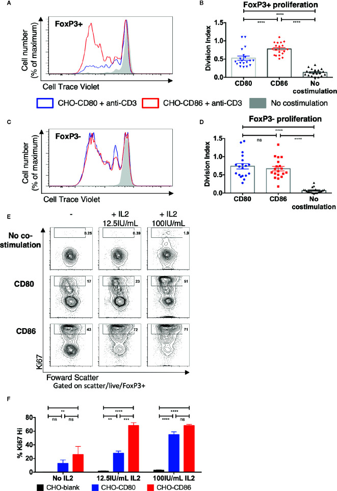
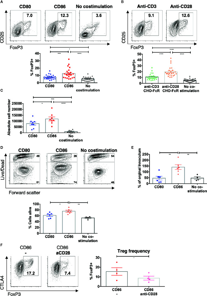
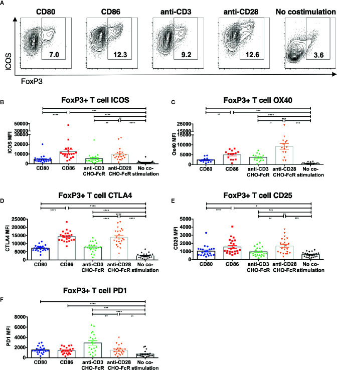
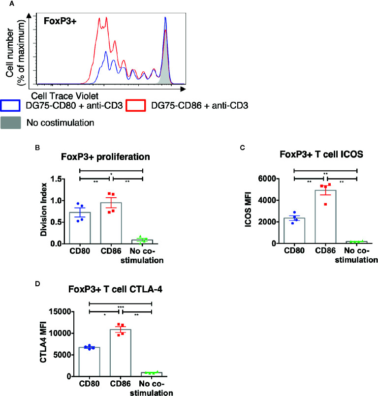
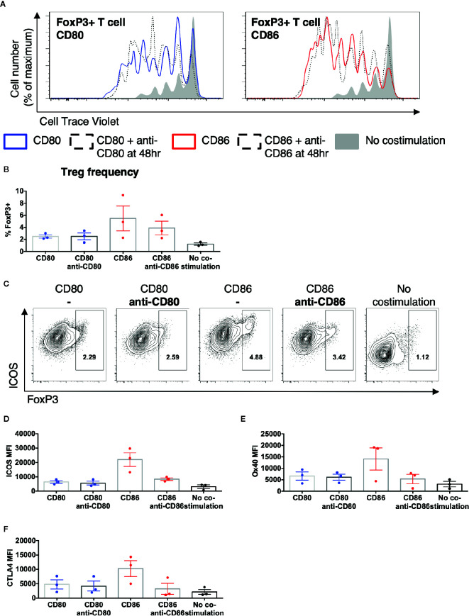
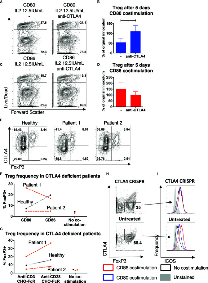
CD80 and CD86 are expressed on antigen presenting cells and are required to engage their shared receptor, CD28, for the costimulation of CD4 T cells. It is unclear why two stimulatory ligands with overlapping roles have evolved. CD80 and CD86 also bind the regulatory molecule CTLA-4. We explored the role of CD80 and CD86 in the homeostasis and proliferation of CD4+FoxP3+ regulatory T cells (Treg), which constitutively express high levels of CTLA-4 yet are critically dependent upon CD28 signals. We observed that CD86 was the dominant ligand for Treg proliferation, survival, and maintenance of a regulatory phenotype, with higher expression of CTLA-4, ICOS, and OX40. We also explored whether CD80-CD28 interactions were specifically compromised by CTLA-4 and found that antibody blockade, clinical deficiency of CTLA-4 and CRISPR-Cas9 deletion of CTLA-4 all improved Treg survival following CD80 stimulation. Taken together, our data suggest that CD86 is the dominant costimulatory ligand for Treg homeostasis, despite its lower affinity for CD28, because CD80-CD28 interactions are selectively impaired by the high levels of CTLA-4. These data suggest a cell intrinsic role for CTLA-4 in regulating CD28 costimulation by direct competition for CD80, and indicate that that CD80 and CD86 have discrete roles in CD28 costimulation of CD4 T cells in the presence of high levels of CTLA-4.

Keywords: CD28; CD80; CD86; CTLA-4; costimulation; homeostasis; regulatory T cells.


Conflict of interest statement

The authors declare that the research was conducted in the absence of any commercial or financial relationships that could be construed as a potential conflict of interest.

Figures

Figure 1
Figure 1
Treg selectively proliferate in response to CD86 costimulation. (A–D) CTV labelled total memory CD4 T cells isolated from healthy donors were cultured for 5 days with CHO-CD80, CHO-CD86, or untransduced CHO (no costimulation), at a ratio of 10 T cells to one CHO cell, with 0.5 μg/ml anti-human-CD3. (A) representative proliferation traces and (B) division indices of FoxP3+ T cells are shown. (C) proliferation traces and (D) division indices of FoxP3- conventional memory CD4 T cells (19 independent experiments). (E, F) purified Treg isolated from healthy donors were cultured with CHO-CD80, CHO-CD86, or untransduced CHO cells, at 10:1 ratio for 5 days, with 0.5 μg/ml anti-human-CD3 with increasing additional IL2. (E) representative Treg Ki67 expression. (F) percentage of Ki76+ Treg after 5 days (three independent experiments). Statistical significance of Tukey’s multiple comparisons tests: ns p>0.05, **p ≤ 0.01, ***p ≤ 0.001, ****p ≤ 0.0001 using One-way analysis of variance (ANOVA) (B, D) and two-way ANOVA (F).
Figure 2
Figure 2
CD86-CD28 interactions drive accumulation of Treg. CTV labelled total memory CD4 T cells isolated from healthy donors were cultured for 5 days with (A) CHO-CD80, CHO-CD86 or untransduced CHO (no costimulation), at a ratio of 10 T cells to one CHO cell, with 0.5 μg/ml anti-human-CD3 or B) CHO-FcR at 2.5:1 ratio with 1 μg/ml anti-human-CD3 or anti-human-CD28. (A, B) Frequency of FoxP3+ amongst divided T cells (24 independent experiments). (C) absolute number of FoxP3+ cells after stimulation (under same conditions as A) (eight independent experiments). Purified Treg isolated from healthy donors were cultured with CHO-CD80, CHO-CD86 or untransduced CHO cells, at 10:1 ratio for 5 days, with 0.5 μg/ml anti-human-CD3 with 12.5 IU/ml IL2. (D) Representative flow cytometry plots demonstrate greater viability of Treg after 5 days with CD86. (E) shows the total number of Treg as a % of the starting population (six independent experiments). (F) CTV labeled total memory CD4 T cells isolated from healthy donors were cultured for 5 days with CHO-CD86 at 10:1 ratio with 0.5 μg/ml anti-human-CD3 and 1 μg/ml anti-human-CD28. Representative plots and graph demonstrate reduced frequency of FoxP3+ cells amongst divided T cells with blockade of CD28 (four independent experiments). One-way ANOVA with Tukey’s multiple comparisons tests (A–D) and paired t-test (E): *p ≤ 0.05, **p ≤ 0.01, ***p ≤ 0.001, ****p ≤ 0.0001.
Figure 3
Figure 3
CD86-CD28 costimulation enhances expression of CD25, CTLA-4, OX40 and ICOS. CTV labelled total memory CD4 T cells isolated from healthy donors were cultured for 5 days with CHO-CD80, CHO-CD86 or untransduced CHO (no costimulation), at a ratio of 10 T cells to one CHO cell, with 0.5 μg/ml anti-human-CD3 or with CHO-FcR at 2.5:1 ratio with 1 μg/ml anti-human-CD3 or anti-human-CD28. After 5 days levels of (A, B) ICOS, (C) OX40, (D) CTLA4, (E) CD25 and (F) PD1 in divided CD4+FoxP3+ T cells were assessed by flow cytometry. (A) representative flow cytometry plots showing ICOS expression. (B–F) aggregate data from 22 independent experiments. One-way ANOVA with Tukey’s multiple comparisons tests: *p ≤ 0.05, **p ≤ 0.01, ***p ≤ 0.001, ****p ≤ 0.0001.
Figure 4
Figure 4
CD86-CD28 costimulation from human model APCs enhances T cell proliferation and expression of CTLA-4 and ICOS. CTV labelled total memory CD4 T cells isolated from healthy donors were cultured for 5 days with DG75-CD80, DG75-CD86, or untransduced DG75 (no costimulation), at a ratio of 1:1 T cells to DG75, with 0.5 μg/ml anti-human-CD3. After 5 days proliferation was assessed by CTV dilution. (A) representative proliferation traces of FoxP3+ cells following costimulation with CD86 or CD80. (B) FoxP3+ T cell division indices, (C) ICOS and (D) CTLA4 expression following costimulation with CD80 or CD86. Data from four independent experiments. One-way ANOVA with Tukey’s multiple comparisons tests: *p ≤ 0.05, **p ≤ 0.01, ***p ≤ 0.001.
Figure 5
Figure 5
Treg phenotype is dependent upon persistent CD86 costimulation. CTV labelled total memory CD4 T cells isolated from healthy donors were cultured for 5 days with CHO-CD80, CHO-CD-86, or untransduced CHO (no costimulation), at a ratio of 10 T cells to one CHO cell, with 0.5 μg/ml anti-human-CD3 and the addition of anti-CD80 or anti-CD86 48 h. (A) representative proliferation traces of FoxP3+ T cells following blockade of CD80 or CD86 after 48 h of stimulation. (B) frequency of FoxP3+ amongst divided CD4 T cells following addition of anti-CD80 or anti-CD86 antibodies after 48 h of culture. (C) representative flow cytometry plots of showing ICOS expression after 5 days in divided CD4 T cells following the addition of anti-CD80 or anti-CD86 antibodies after 48 h of culture. Aggregate data for the expression of (D) ICOS, (E) OX40, (F) CTLA4 in divided CD4+FoxP3+ve T cells. Three independent experiments.
Figure 6
Figure 6
CTLA4 prevents Treg from receiving costimulation from CD80. Purified Treg isolated from healthy donors were cultured with CHO-CD80, CHO-D86, at 10:1 ratio for 5 days, with 0.5 μg/ml anti-human-CD3 and 12.5 IU/ml IL2 with anti-CTLA4 antibody. (A, B) cytometry plots demonstrate survival of Treg following costimulation with CD80 with- and without anti-CTLA4 antibodies and (C, D) similarly with CD86 costimulation. Representative plots and aggregate data from four independent experiments. (E) Total memory CD4 T cells were isolated from a healthy donor, a patient with LRBA deficiency (patient 1) and a patient CTLA4 deficiency (patient 2). T cells were stimulated for 16 h with 2:1 T cells: anti-CD3 and anti-CD28 coated activator beads and CTLA4 and FoxP3 expression assessed by flow cytometry, demonstrating CTLA4 deficiency in both CD4+FoxP3+ and CD4+FoxP3- T cells. (F, G) CTV labeled total memory CD4 T cells, isolated from patients 1 and 2 and a healthy donor, were stimulated at 10:1 ratio with CHO-CD80, CHO-CD86, or untransduced CHO (no costimulation) with 0.5 μg/ml anti-human-CD3 (F), or at 2.5:1 with CHO-FcR and 1 μg/ml anti-human-CD3 or anti-human-CD28 antibodies (G) for 5 days. Graphs illustrate the frequency of FoxP3+ cells among the divided CD4 T cells. (H, I) Treg were purified from healthy donors and underwent CTLA-4 deletion by CRISPR and were then cultured 10:1 with CHO-CD80, CHO-CD86, or untransduced CHO (no costimulation) and 0.5 μg/ml anti-human-CD3 for 5 days. (H) demonstrates CTLA4 levels in CRISPR/Cas9 treated and untreated cells after 5 days costimulation. (I) Histograms demonstrating ICOS levels following either CD86 or CD80 costimulation of CTLA4-deleted or untreated, CTLA4-replete Treg (representative plot of two independent experiments). Paired t-test: *p ≤ 0.05.

Similar articles

Cited by

References

    1. Viola A, Lanzavecchia A. T cell activation determined by T cell receptor number and tunable thresholds. Science (1996) 273(5271):104–6. 10.1126/science.273.5271.104 - DOI - PubMed
    1. Harding FA, McArthur JG, Gross JA, Raulet DH, Allison JP. CD28-mediated signalling co-stimulates murine T cells and prevents induction of anergy in T-cell clones. Nature (1992) 356(6370):607–9. 10.1038/356607a0 - DOI - PubMed
    1. Esensten JH, Helou YA, Chopra G, Weiss A, Bluestone JA. CD28 Costimulation: From Mechanism to Therapy. Immunity (2016) 44(5):973–88. 10.1016/j.immuni.2016.04.020 - DOI - PMC - PubMed
    1. Hansen JD, Du Pasquier L, Lefranc MP, Lopez V, Benmansour A, Boudinot P. The B7 family of immunoregulatory receptors: a comparative and evolutionary perspective. Mol Immunol (2009) 46(3):457–72. 10.1016/j.molimm.2008.10.007 - DOI - PubMed
    1. Collins M, Ling V, Carreno BM. The B7 family of immune-regulatory ligands. Genome Biol (2005) 6(6):223. 10.1186/gb-2005-6-6-223 - DOI - PMC - PubMed

Publication types