

Case Reports
doi: 10.1016/j.radcr.2020.12.014.
eCollection 2021 Mar.
Myositis and myopathy of sarcoidosis: A case report
Affiliations
- PMID: 33363677
- PMCID: PMC7753091
- DOI: 10.1016/j.radcr.2020.12.014
Item in Clipboard
Case Reports
Myositis and myopathy of sarcoidosis: A case report
Radiol Case Rep.
.
Abstract
Muscular manifestations of sarcoidosis are commonly found on biopsy but rare on correlated imaging. We present a rare case of a 36-year-old male patient with sarcoid myositis and image findings of active myositis in the lower back and pelvic girdle musculature. This case suggests considering sarcoidosis as a differential diagnosis in the setting of chest findings and new lower back and lower extremity weakness.
Keywords: Myopathy; Myositis; Sarcoidosis.
© 2020 Published by Elsevier Inc. on behalf of University of Washington.
Figures

Coronal noncontrast CT shows prominent calcified mediastinal and hilar adenopathy (black arrowheads). There is additional prominent bronchiectasis (straight arrows).

Axial noncontrast CT reveals an enlarged main pulmonary artery measuring 3.4 cm (straight white arrows). Marked calcified mediastinal and hilar lymph nodes are present (black arrowheads).

Coronal noncontrast CT with lung windows shows signs of pulmonary fibrosis. There is upper lobe predominant architectural distortion, subpleural interstitial nodularity (black arrowheads), and bronchiectasis with peribronchovascular honeycombing (straight arrows).

Axial CT image. Multifocal patchy edema and atrophy within bilateral pelvic girdle musculature including gluteus maximus (curved arrows), piriformis (white arrowheads), gluteus minimus and medius (black arrowheads), and minimally within iliopsoas (straight arrows) and piriformis. Calcified iliac lymph nodes are also visible.
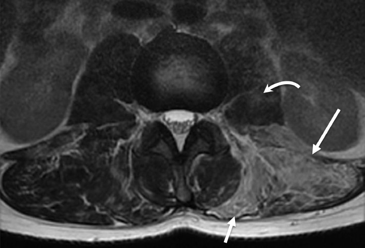
T2-weighted MRI of the lumbar spine reveals edema and atrophy most prominent within the left paraspinous muscles (straight arrows) as well as likely mild edema within the left psoas muscle (curved arrow).

T1-weighted MRI with contrast. Left paraspinous musculature showed enhancement with contrast administration (straight arrows).
References
Publication types
LinkOut - more resources
Full Text Sources
Other Literature Sources
