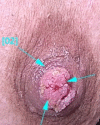
Dermoscopy of nipple adenoma
- PMID: 33363915
- PMCID: PMC7752568
- DOI: 10.1002/ccr3.3398
Dermoscopy of nipple adenoma
Abstract
Characteristic finding of nipple adenoma (NA) in dermoscopy (red dots in linear, radial, or semicircular patterns) can help in accurate clinicopathologic diagnosis of NA vs other inflammatory, benign, and especially malignant nipple lesions.
Keywords: breast; dermoscopy; nipple; nipple adenoma.
© 2020 The Authors. Clinical Case Reports published by John Wiley & Sons Ltd.
Conflict of interest statement
The authors have no conflict of interest to declare.
Figures


References
-
- Cosechen MS, Wojcik ASdL, Piva FM, et al. Erosive adenomatosis of the nipple. An Bras Dermatol. 2011;86(4):17‐20. - PubMed
-
- Spohn GP, Trotter SC, Tozbikian G, et al. Nipple adenoma in a female patient presenting with persistent erythema of the right nipple skin: case report, review of the literature, clinical implications, and relevancy to health care providers who evaluate and treat patients with dermatologic conditions of the breast skin. BMC Dermatol. 2016;16(1):4. - PMC - PubMed
-
- Takashima S, Fujita Y, Miyauchi T, et al. Dermoscopic observation in adenoma of the nipple. J Dermatol. 2015;42(3):341‐342. - PubMed
Publication types
LinkOut - more resources
Full Text Sources

