Review
. 2020 Dec 16:8:100311.
doi: 10.1016/j.ejro.2020.100311. eCollection 2021.

Connective tissue disease-related interstitial lung disease (CTD-ILD) and interstitial lung abnormality (ILA): Evolving concept of CT findings, pathology and management

Affiliations
Review

Connective tissue disease-related interstitial lung disease (CTD-ILD) and interstitial lung abnormality (ILA): Evolving concept of CT findings, pathology and management

Hongseok Yoo et al. Eur J Radiol Open. .

Retraction in

Abstract

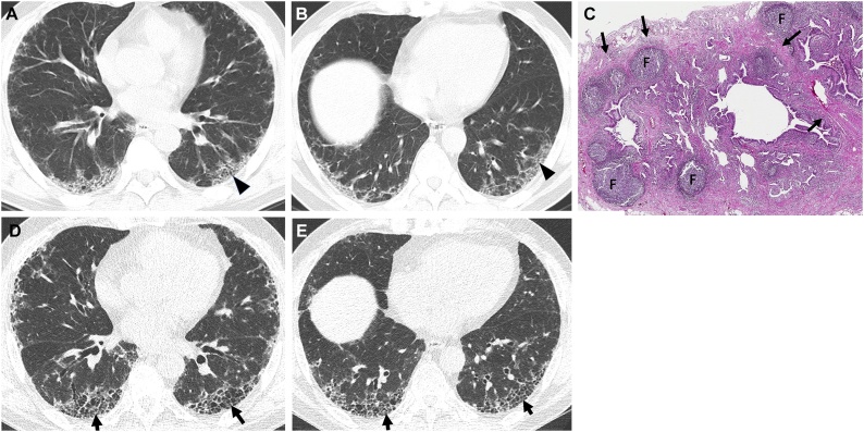
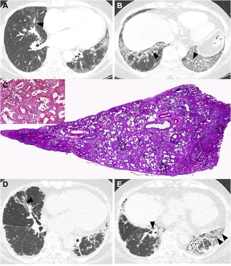
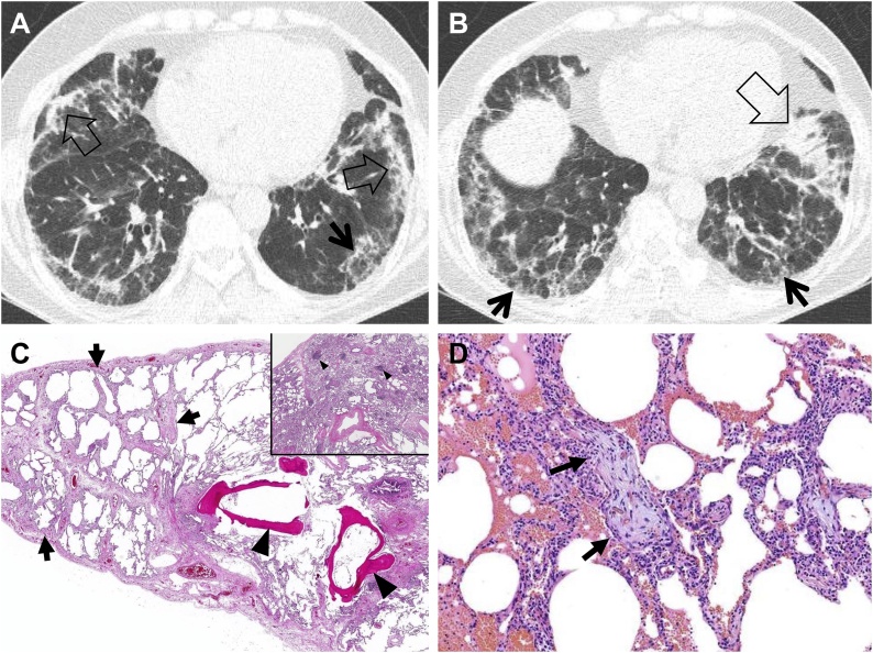
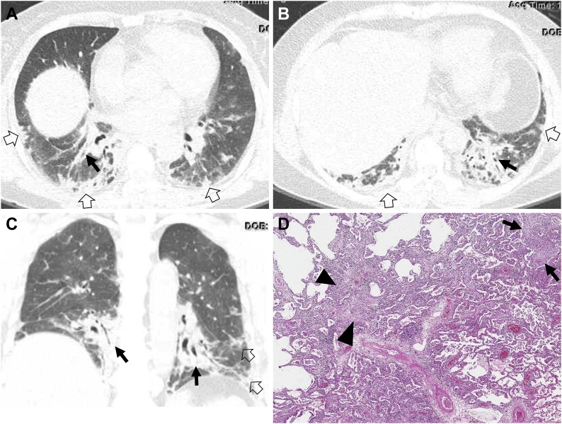
The connective tissue diseases (CTDs) demonstrating features of interstitial lung disease (ILD) include systemic lupus erythematosus (SLE), rheumatoid arthritis (RA), progressive systemic sclerosis (PSS), dermatomyositis (DM) and polymyositis (PM), ankylosing spondylitis (AS), Sjogren's syndrome (SS), and mixed connective tissue disease (MCTD). In RA patients in particular, interstitial lung abnormality (ILA) (of varying degrees; severe vs. mild) is reported to occur in approximately 20-60 % of individuals and CT disease progression occurs in approximately 35-45 % of them. The ILAs have been associated with a spectrum of functional and physiologic decrement. The identification of progressive ILA may enable appropriate surveillance and the commencement of treatment with the goal of improving morbidity and mortality rates of established RA-ILD. Subpleural distribution and higher baseline ILA/ILD extent were risk factors associated with disease progression. At histopathologic analysis, connective tissue disease-related interstitial lung diseases (CTD-ILDs) are diverse and include nonspecific interstitial pneumonia (NSIP), usual interstitial pneumonia (UIP), organizing pneumonia (OP), apical fibrosis, diffuse alveolar damage (DAD), and lymphoid interstitial pneumonia (LIP). Even though proportions of ILDs vary, NSIP pattern accounts for a large proportion, especially in PSS, DM/PM and MCTD, followed by UIP pattern. Evidence has been published that treatment of subclinical CT lung abnormalities showing a tendency to progress to ILD may stabilize the CT alterations. The identification of subclinical lung abnormalities can be appropriate in the management of the disease and CT appears to be the gold standard for the evaluation of lung parenchyma.

Keywords: CTD, Connective tissue disease; CTD-ILD, (Connective Tissue Disease-Related Interstitial Lung Disease); Connective tissue disease; DM, Dermatomyositis; IIP, Idiopathic interstitial pneumonia; ILA, Interstitial lung abnormality; ILD, Interstitial lung disease; IPAF, Interstitial pneumonitis with autoimmune features; IPF, Idiopathic pulmonary fibrosis; Interstitial lung abnormality; Interstitial lung disease; MCTD, Mixed connective tissue disease; NSIP, Nonspecific Interstitial Pneumonia; OP, Organizing pneumonia; PM, Polymyositis; PSS, Progressive Systemic Sclerosis; RA, Rheumatoid Arthritis; SLE, Systemic Lupus Erythematosus; SS, Sjogren’s Syndrome; UCTD, Undifferentiated Connective Tissue Disease; UIP, Usual Interstitial Pneumonia.


Conflict of interest statement

The authors report no declarations of interest.

Figures

Fig. 1
Fig. 1
Interstitial pneumonia with autoimmune features (IPAF) in a 67-year-old man. (a, b) Lung window images of CT scans obtained at levels of right inferior pulmonary vein (a) and liver dome (b), respectively, show subpleural reticulation and traction bronchiolectasis (arrowheads) in both lungs. Patient had positive serologic tests; fluorescent antinuclear antibody (FANA, 1:320) and perinuclear antineutrophil cytoplasmic antibody (pANCA, 1:320). (c) Low-power magnification of lung demonstrates collapse of two secondary lobules (arrows) resulting in dilation of distal small airways (so-called honeycombing) with super imposed lymphoid follicles with reactive germinal centers (F). The histologic pattern is most consistent with UIP, the superimposed lymphoid follicles with reactive germinal centers suggest CTD as the underlying cause of all the histopathologic findings but are not specific. (d, e). Four-year follow-up CT scans obtained at similar levels to a & b, respectively, depict apparent areas of CT honeycombing (arrows) in posterior aspects of both lower lobes.
Fig. 2
Fig. 2
Nonspecific interstitial pneumonia in a 48-year-old woman with dermatomyositis. (a, b) Lung window images of CT scans obtained at levels of right inferior pulmonary vein (a) and liver dome (b), respectively, show reticulation and traction bronchiectasis (arrowheads) in both lungs with lower lung zone predominance. (c) Low power magnification, and medium power magnification (inset) of lung demonstrates temporally uniform diffuse parenchymal fibrosis typical of fibrotic NSIP. Superimposed are diffusely scattered lymphoid follicles containing reactive germinal centers, which suggest CTD as the underlying cause of the fibrotic NSIP but are not specific. (d, e). Ten-year follow-up CT scans obtained at similar levels to a & b, respectively, depict progression of pulmonary fibrosis with apparent areas of traction bronchiectasis in both lungs.
Fig. 3
Fig. 3
Fibrosing organizing pneumonia in a 58-year-old woman with interstitial pneumonia with autoimmune features (IPAF; antineutrophil antibody [ANA], 1:160 and morning stiffness). (a, b) Lung window images of CT scans obtained at levels of cardiac ventricle (a) and liver dome (b), respectively, show patchy distribution of mixed areas of band-like consolidation (open arrows) and reticulation (arrows) in both lungs. (c) Low power magnification of lung demonstrates temporally uniform diffuse lung fibrosis typical of fibrotic NSIP (arrows) associated with dendritic ossification (arrowheads), which is a reflection of chronicity of injury. Inset: Lymphoid follicles containing reactive germinal centers (arrowheads) are randomly scattered in the lung parenchyma suggesting CTD as the underlying cause of the fibrotic NSIP but are not specific. (d) Medium power magnification of lung from the right middle lobe demonstrates organizing pneumonia characterized by airspaces plugs of loose myxoid fibrous tissue (arrows). Organizing pneumonia is a non-specific histologic finding and is one of the lungs most common reactions to injury from any cause.
Fig. 4
Fig. 4
Organizing pneumonia in a 56-year-old woman with dermatomyositis. (a, b) Lung window images of CT scans obtained at levels of liver dome (a) and 3 cm inferior to a (b), respectively, show patchy distribution of consolidation along bronchovascular bundles (arrows) and subpleural lungs (open arrows) in both lungs. (c) Coronal reformatted image demonstrates consolidation along bronchovascular bundle (arrows) and subpleural (open arrows) lungs. (d) Low power magnification of lung demonstrating organizing pneumonia (arrows) transitioning to fibrotic NSIP (arrowheads). CTD is in the etiologic differential of fibrotic NSIP, but there are no histologic findings in this image that suggest CTD as the underlying cause in contrast to the lymphoid follicles with reactive germinal centers seen in Fig. 1, Fig. 2.
Fig. 5
Fig. 5
Connective tissue disease-related pulmonary fibrosis showing straight-edge sign. (a) Coronal reformatted image in a 49-year-old woman with systemic sclerosis shows pulmonary fibrosis composed of reticulation and ground-glass opacity confined to lower lung zone with straight-edge (arrows) sign. (b) Coronal reformatted image in a 31-year-old Sjogren’s syndrome demonstrates reticulation and honeycombing in lower lung zones with straight-edge (arrows) sign.
Fig. 6
Fig. 6
Connective tissue disease-related pulmonary fibrosis showing exuberant honeycombing sign in a 57-year-old woman with rheumatoid arthritis. (a, b) Lung window images of CT scans obtained at levels of right inferior pulmonary vein (a) and suprahepatic inferior vena cava (b), respectively, show extensive honeycombing in lower lung zones. Also note pericardial effusion (PE) and cardiomegaly.
Fig. 7
Fig. 7
Connective tissue disease-related pulmonary fibrosis showing anterior upper lobe sign in a 65-year-old woman with rheumatoid arthritis. (a) Lung window image of CT scan obtained at level of aortic arch demonstrates large area of honeycombing (arrows) in anterior aspects of both upper lobes. (b) Sagittal reformatted image discloses reticular lesions in anterior upper lobe (arrows) in left side. Also note reticular lesions in subpleural areas (arrowheads) of left lower lung zones.
Fig. 8
Fig. 8
Acute exacerbation of usual interstitial pneumonia in a 49-year-old man with rheumatoid arthritis. (a, b) Lung window images of CT scans obtained at levels of right inferior pulmonary vein (a) and liver dome (b), respectively, show extensive honeycombing in middle and lower lung zones. Also note large areas of mixed consolidation and ground-glass opacity (arrowheads) in a. (c, d) Coronal reformatted images demonstrate patchy and large areas of ground-glass opacity with crazy-paving appearance (arrows) and consolidation (open arrows). Also note large areas of honeycombing as underlying disease.
Fig. 9
Fig. 9
Interstitial lung abnormality and lung squamous cell carcinoma in an 80-year-old man with rheumatoid arthritis. (a) Lung window image of CT scan obtained at level of liver dome shows subpleural fibrotic interstitial lung abnormality composed of reticulation and traction bronchiolectasis (arrowheads). Also note a 22-mm-sized nodule (open arrow) in left lung base. (b) Fused CT/PET image demonstrate high FDG uptake within left lower lung zone tumor (proved to be squamous cell carcinoma). (c) Low power magnification of lung demonstrating cystic dilation of distal small airways with rare fibroblast foci in the walls of the cysts consistent with UIP that are associated with patchy, moderate lymphoid infiltrates.

References

    1. American Thoracic Society, European Respiratory Society, American Thoracic Society/European Respiratory Society International Multidisciplinary Consensus Classification of the Idiopathic Interstitial Pneumonias This joint statement of the American Thoracic Society (ATS), and the European Respiratory Society (ERS) was adopted by the ATS board of directors, June 2001 and by the ERS Executive Committee, June 2001. Am. J. Respir. Crit. Care Med. 2002;165:277–304. - PubMed
    1. Lederer D.J., Martinez F.J. Idiopathic pulmonary fibrosis. N. Engl. J. Med. 2018;378:1811–1823. - PubMed
    1. Jeganathan N., Sathananthan M. Connective tissue disease-related interstitial lung disease: prevalence, patterns, predictors, prognosis, and treatment. Lung. 2020;198:735–759. - PubMed
    1. Ng K.H., Chen D.Y., Lin C.H., Chao W.C., Chen Y.M., Chen Y.H., Huang W.N., Hsieh T.Y., Lai K.L., Tang K.T., Chen H.H. Risk of interstitial lung disease in patients with newly diagnosed systemic autoimmune rheumatic disease: a nationwide, population-based cohort study. Semin. Arthritis Rheum. 2020;50:840–845. - PubMed
    1. Kim E.A., Lee K.S., Johkoh T., Kim T.S., Suh G.Y., Kwon O.J., Han J. Interstitial lung diseases associated with collagen vascular diseases: radiologic and histopathologic findings. Radiographics. 2002;22:S151–165. Spec No. - PubMed