. 2020 Dec 21;5(1):e513.
doi: 10.1097/HS9.0000000000000513. eCollection 2021 Jan.

Recurrent NR3C1 Aberrations at First Diagnosis Relate to Steroid Resistance in Pediatric T-Cell Acute Lymphoblastic Leukemia Patients

Affiliations

Recurrent NR3C1 Aberrations at First Diagnosis Relate to Steroid Resistance in Pediatric T-Cell Acute Lymphoblastic Leukemia Patients

Jordy C G van der Zwet et al. Hemasphere. .

Abstract

The glucocorticoid receptor NR3C1 is essential for steroid-induced apoptosis, and deletions of this gene have been recurrently identified at disease relapse for acute lymphoblastic leukemia (ALL) patients. Here, we demonstrate that recurrent NR3C1 inactivating aberrations-including deletions, missense, and nonsense mutations-are identified in 7% of pediatric T-cell ALL patients at diagnosis. These aberrations are frequently present in early thymic progenitor-ALL patients and relate to steroid resistance. Functional modeling of NR3C1 aberrations in pre-B ALL and T-cell ALL cell lines demonstrate that aberrations decreasing NR3C1 expression are important contributors to steroid resistance at disease diagnosis. Relative NR3C1 messenger RNA expression in primary diagnostic patient samples, however, does not correlate with steroid response.


Conflict of interest statement

The authors declare no competing interest.

Figures

Figure 1.
Figure 1.
Schematic overview of the NR3C1 protein. Missense mutations (blue) and frameshift (red) mutations, as found in our cohort of 146 T-ALL patients, have been indicated. T-ALL = T-cell acute lymphoblastic leukemia.
Figure 2.
Figure 2.
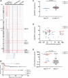
Characteristics and steroid response of NR3C1 aberrations in primary T-ALL patient samples. (A), Array-CGH data of 92 diagnostic biopsies from pediatric T-ALL patients. Patients are horizontally orientated and T-ALL subtypes are indicated as determined by gene expression profiling. Deleted regions of the 5 NR3C1-deleted patients detected by Array-CGH are characterized by the larger blue lesions around the 5q31 locus. (B), Kaplan-Meier survival analysis of 146 T-ALL patients who have been treated with different treatment protocols (Supplemental Table 1, http://links.lww.com/HS/A123). The survival for either NR3C1 deleted or NR3C1 mutated patients was compared to patients that lack NR3C1 aberrations and analyzed using the log-rank (Mantel-Cox) test. (C), Median NR3C1 expression of 116 diagnostic biopsies from pediatric T-ALL patients determined by U133plus 2 arrays. NR3C1 deleted cases (blue dots) had significant lower NR3C1 expression levels compared to non-NR3C1 deleted cases (including NR3C1 mutated patients highlighted by red triangles) (Mann-Whitney U test, P = 0.0017). (D), Matched NR3C1 expression data to in vitro prednisolone response of 83 diagnostic biopsies from pediatric T-ALL patients. (E), In vitro prednisolone response of 96 diagnostic biopsies from pediatric T-ALL patients. Patients that harbor NR3C1 aberrations (deletion [blue dots] or mutation [red triangles]) were significantly more resistant to prednisolone compared to NR3C1 wild-type patients (Mann-Whitney U test, P = 0.0078). CGH = array-based comparative genomic hybridization; ETP-ALL = early thymic progenitor acute lymphoblastic leukemia; T-ALL = T-cell acute lymphoblastic leukemia.
Figure 3.
Figure 3.
NR3C1 expression levels predict steroid response in REH cells. (A), Steroid response curves for doxycycline-induced (+dox) or noninduced (–dox) steroid-resistant REH cells following 96 h exposure to serial dilutions of prednisolone. Cell survival was determined by flow cytometry measured cell counts. REH cells were transfected with a doxycycline-inducible NR3C1 wild-type construct. (B) and (C), Steroid response curves and NR3C1 protein levels and steroid response curves of NR3C1-REH cells (eg, doxycycline-induced REH NR3C1 wild-type cells) that have been transduced with NR3C1-directed lentiviral shRNA constructs (sh1-4). Control shRNA construct sh5 is directed against the 3′UTR of NR3C1, which is not included in the NR3C1 expression construct. (D) and (E), Steroid response curves and (endogenous or short hairpin reduced) NR3C1 protein levels of steroid-sensitive SUP-T1 cells. shRNA constructs were induced by IPTG 4 d or 1 d prior to—or at the start (day 0) of steroid treatment. Steroid response was determined after 96 h by flow cytometry measured cell counts. IPTG = isopropyl β- d-1-thiogalactopyranoside; shRNA = short hairpin RNA.
Figure 4.
Figure 4.
Nontruncated NR3C1 missense mutations induce efficient steroid-induced cell death. (A), Western blot results of total NR3C1 levels for REH cell lines transfected with doxycycline-inducible NR3C1 or mutant NR3C1-constructs as indicated. (B), Transcriptional steroid response of REH cells that have been transfected with doxycycline-inducible WT or mutant NR3C1 constructs. Doxycycline-induced cells were treated with 250 μg/mL prednisolone. Expression of glucocorticoid receptor target genes GILZ, FKBP5, and SGK1 was determined at 8, 24, and 48 h following prednisolone treatment. (C), Steroid response curves of REH parental and REH cells transfected with doxycycline-inducible mutant NR3C1 constructs. GAPDH = glyceraldehyde 3-phosphate dehydrogenase; WT = wild-type.

Similar articles

Cited by

References

    1. Riehm H, Reiter A, Schrappe M, et al. [Corticosteroid-dependent reduction of leukocyte count in blood as a prognostic factor in acute lymphoblastic leukemia in childhood (therapy study ALL-BFM 83)]. Klin Padiatr. 1987; 199:151–160. - PubMed
    1. Griffin TC, Shuster JJ, Buchanan GR, et al. Slow disappearance of peripheral blood blasts is an adverse prognostic factor in childhood T cell acute lymphoblastic leukemia: a Pediatric Oncology Group study. Leukemia. 2000; 14:792–795. - PubMed
    1. Weikum ER, Knuesel MT, Ortlund EA, et al. Glucocorticoid receptor control of transcription: precision and plasticity via allostery. Nat Rev Mol Cell Biol. 2017; 18:159–174. - PMC - PubMed
    1. Wandler AM, Huang BJ, Craig JW, et al. Loss of glucocorticoid receptor expression mediates in vivo dexamethasone resistance in T-cell acute lymphoblastic leukemia. Leukemia. 2020; 34:2025–2037. - PMC - PubMed
    1. Miller AL, Garza AS, Johnson BH, et al. Pathway interactions between MAPKs, mTOR, PKA, and the glucocorticoid receptor in lymphoid cells. Cancer Cell Int. 2007; 7:3. - PMC - PubMed