. 2020 Dec 7:11:593468.
doi: 10.3389/fphys.2020.593468. eCollection 2020.

The Acute Effects of 5 Fluorouracil on Skeletal Muscle Resident and Infiltrating Immune Cells in Mice

Affiliations

The Acute Effects of 5 Fluorouracil on Skeletal Muscle Resident and Infiltrating Immune Cells in Mice

Brandon N VanderVeen et al. Front Physiol. .

Abstract

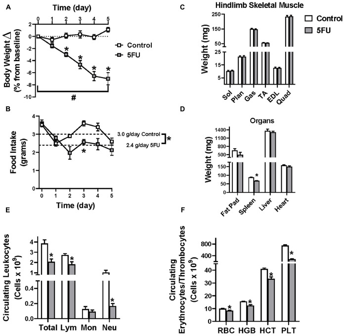
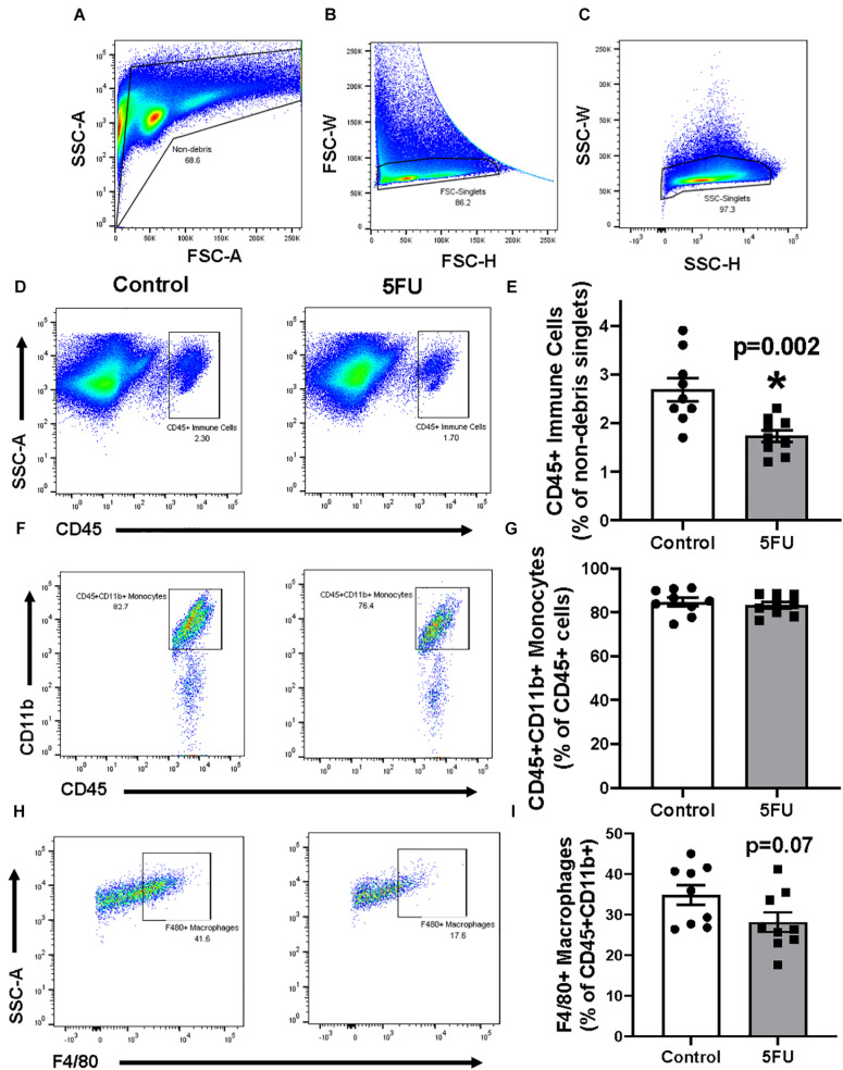
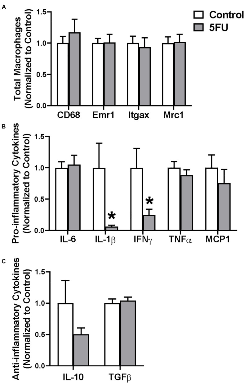
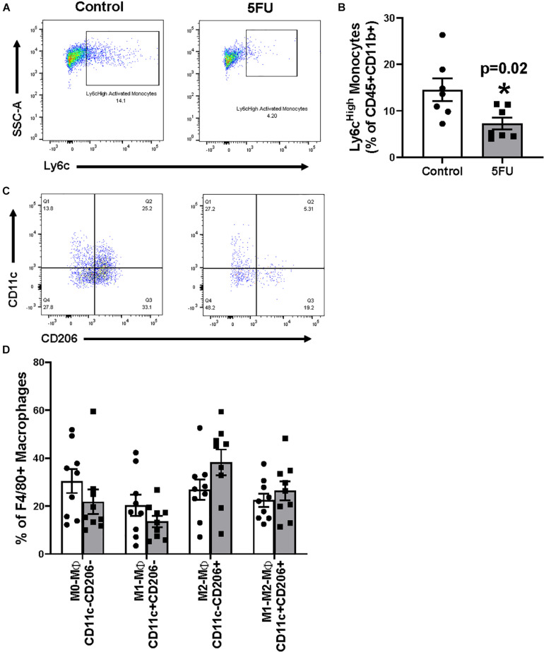
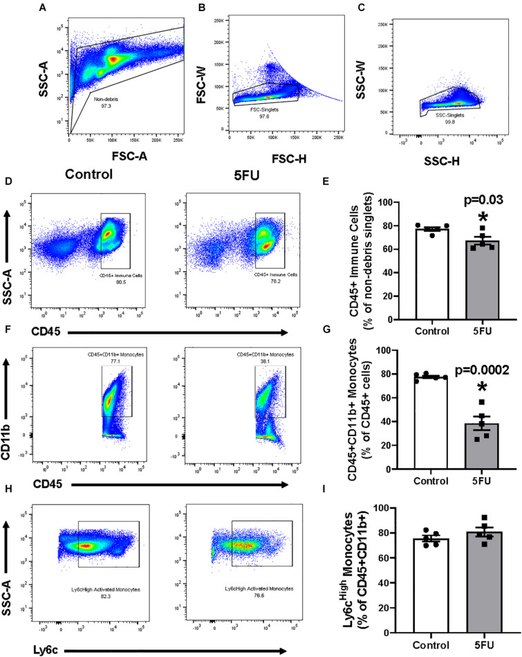
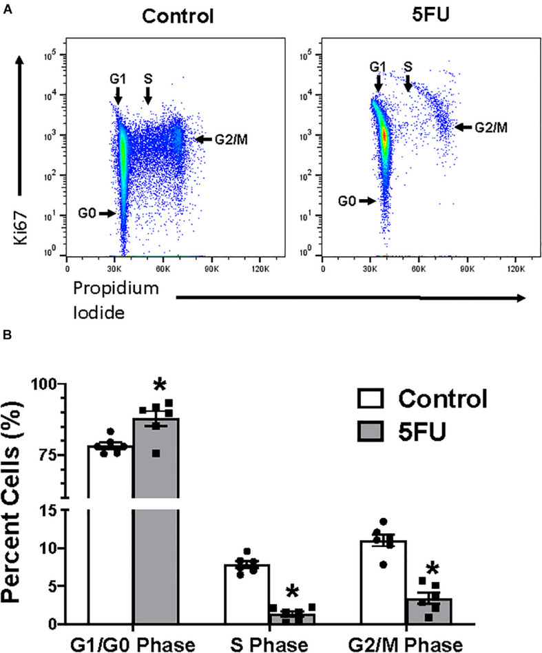
5 fluorouracil (5FU) has been a first-choice chemotherapy drug for several cancer types (e.g., colon, breast, head, and neck); however, its efficacy is diminished by patient acquired resistance and pervasive side effects. Leukopenia is a hallmark of 5FU; however, the impact of 5FU-induced leukopenia on healthy tissue is only becoming unearthed. Recently, skeletal muscle has been shown to be impacted by 5FU in clinical and preclinical settings and weakness and fatigue remain among the most consistent complaints in cancer patients undergoing chemotherapy. Monocytes, or more specifically macrophages, are the predominate immune cell in skeletal muscle which regulate turnover and homeostasis through removal of damaged or old materials as well as coordinate skeletal muscle repair and remodeling. Whether 5FU-induced leukopenia extends beyond circulation to impact resident and infiltrating skeletal muscle immune cells has not been examined. The purpose of the study was to examine the acute effects of 5FU on resident and infiltrating skeletal muscle monocytes and inflammatory mediators. Male C57BL/6 mice were given a physiologically translatable dose (35 mg/kg) of 5FU, or PBS, i.p. once daily for 5 days to recapitulate 1 dosing cycle. Our results demonstrate that 5FU reduced circulating leukocytes, erythrocytes, and thrombocytes while inducing significant body weight loss (>5%). Flow cytometry analysis of the skeletal muscle indicated a reduction in total CD45+ immune cells with a corresponding decrease in total CD45+CD11b+ monocytes. There was a strong relationship between circulating leukocytes and skeletal muscle CD45+ immune cells. Skeletal muscle Ly6cHigh activated monocytes and M1-like macrophages were reduced with 5FU treatment while total M2-like CD206+CD11c- macrophages were unchanged. Interestingly, 5FU reduced bone marrow CD45+ immune cells and CD45+CD11b+ monocytes. Our results demonstrate that 5FU induced body weight loss and decreased skeletal muscle CD45+ immune cells in association with a reduction in infiltrating Ly6cHigh monocytes. Interestingly, the loss of skeletal muscle immune cells occurred with bone marrow cell cycle arrest. Together our results highlight that skeletal muscle is sensitive to 5FU's off-target effects which disrupts both circulating and skeletal muscle immune cells.

Keywords: bone marrow; chemotherapy; macrophages; monocytes; skeletal muscle.


Conflict of interest statement

BV, DF, and EM were employed by the company AcePre LLC. The remaining authors declare that the research was conducted in the absence of any commercial or financial relationships that could be construed as a potential conflict of interest.

Figures

FIGURE 1
FIGURE 1
Animal characteristics. 5-fluorouracil (5FU) was solubilized in phosphate buffered saline at 3.5 mg/mL and administered to the mice at 35 mg/kg via intraperitoneal injection once daily for 5 days. (A) Relative body weight change shown as the % change from day 0 throughout the duration of the study. (B) Daily food intake in grams throughout the duration of the study. Dotted line illustrates the average daily food intake in grams (g) per day over the course of the 5 days of treatment. (C) Select hindlimb muscle weights given in milligrams (mg) after 5 days of 5FU. (D) Select organ weights in mg after 5 days of 5FU. (E) Circulating leukocytes given as # of cells × 109/L after 5 days of 5FU. (F) Circulating erythrocytes and thrombocytes given as # of cells × 109/L after 5 days of 5FU. Sol, soleus; Plan, plantaris; Gas, gastrocnemius; TA, tibialis anterior; EDL, extensor digitorum longus; Quad, quadriceps; Lym, lymphocytes; Mon, monocytes; Neu, neutrophils; RBC, red blood cells; HBG, hemoglobin; HCT, hematocrit; PLT, platelets. Significance was set at p < 0.05. *Significantly different from Control using a student’s t-test. #Significantly different from Day 0 using a repeated measures Two-way ANOVA.
FIGURE 2
FIGURE 2
The effects of 5FU on skeletal muscle immune cells. (A) Cells were gated for non-debris (SSC-A × FSC-A), (B) FSC singlets (FSC-W × FSC-H), (C) and SSC singlets (SSC-W × SSC-H; A-right). (D) Non-debris singlet cells were then gated for total immune cells with CD45+. (E) CD45+ cells were quantified and shown in the bar graph as the relative % of non-debris singlets. (F) CD45+ cells were gated for monocytes with CD11b+. (G) CD45+CD11b+ cells were quantified and shown in the bar graph as the relative % of CD45+ cells. (H) CD45+CD11b+ were then gated for macrophages with F4/80. (I) F4/80+ cells were quantified and shown in the bar graph as the relative % of CD45+CD11b+ cells. Significance was set at p < 0.05. *Significantly different from Control using a student’s t-test.
FIGURE 3
FIGURE 3
The effects of 5FU on skeletal muscle macrophage gene expression. (A) Relative gene expression of total macrophage genes, CD68 and Emr1 (F4/80), M1-like macrophage gene, Itgax (CD11c), and M2-like macrophage gene, Mrc1 (CD206). (B) Relative gene expression of pro-inflammatory genes, Interleukin (IL) 6, IL-1β, Interferon (IFN) γ, Tumor necrosis factor (TNF) α, and monocyte chemoattractant protein (MCP) 1. (C) Relative gene expression of anti-inflammatory genes IL-10 and transforming growth factor (TGF) β. Significance was set at p < 0.05. *Significantly different from Control using a student’s t-test.
FIGURE 4
FIGURE 4
The effects of 5FU on infiltrating skeletal muscle monocytes and macrophages. (A) CD11b+ monocytes were gated for their activation status using Ly6C. Cells were considered either resident (Ly6cLow) or activated/infiltrating (Ly6cHigh). (B) Ly6cHigh monocytes were quantified and shown in the bar graph as relative % of CD45+CD11b+ cells. (C) F4/80+ macrophages were gated analyzed for their polarization status using CD11c and CD206. (D) CD11c-CD206- cells were considered M0-like macrophages, CD11c+CD206- cells were considered M1-like macrophages, CD11c-CD206+ cells were considered M2-like macrophages, and CD11c+CD206+ cells were considered M1-M2-like transitional macrophages and graphed as the relative % of F480+ macrophages. Significance was set at p < 0.05.*Significantly different from control (t-test).
FIGURE 5
FIGURE 5
The effects of 5FU on bone marrow immune cells. (A) Cells were gated for non-debris (SSC-A × FSC-A), (B) FSC singlets (FSC-W × FSC-H), (C) and SSC singlets (SSC-W × SSC-H; A-right). (D) Non-debris singlet cells were then gated for total immune cells with CD45+. (E) CD45+ cells were quantified and shown in the bar graph as the relative % of non-debris singlets. (F) CD45+ cells were gated for monocytes with CD11b+. (G) CD45+CD11b+ cells were quantified and shown in the bar graph as the relative % of CD45+ cells. (H) CD11b+ monocytes were gated for their activation status using Ly6C. Cells were considered either resident (Ly6cLow) or activated/infiltrating (Ly6cHigh). (I) Ly6cHigh monocytes were quantified and shown in the bar graph as relative % of CD45+CD11b+ cells. Significance was set at p < 0.05. *Significantly different from Control using a student’s t-test.
FIGURE 6
FIGURE 6
The effects of 5FU on bone marrow cell cycle. (A) Cells were fixed and stained with Ki67 and propidium iodide (PI). (B) Cells in the G1/G0, S, and G2/M phases were quantified and shown in the bar graph as the relative % of total cells. Significance was set at p < 0.05.*Significantly different from control (t-test).

References

    1. Abraham J. E., Hiller L., Dorling L., Vallier A. L., Dunn J., Bowden S., et al. (2015). A nested cohort study of 6,248 early breast cancer patients treated in neoadjuvant and adjuvant chemotherapy trials investigating the prognostic value of chemotherapy-related toxicities. BMC Med. 13:306. 10.1186/s12916-015-0547-5 - DOI - PMC - PubMed
    1. Arnold L., Henry A., Poron F., Baba-Amer Y., van Rooijen N., Plonquet A., et al. (2007). Inflammatory monocytes recruited after skeletal muscle injury switch into antiinflammatory macrophages to support myogenesis. J. Exp. Med. 204 1057–1069. - PMC - PubMed
    1. Baechler S., Hobbs R. F., Jacene H. A., Bochud F. O., Wahl R. L., Sgouros G. (2010). Predicting hematologic toxicity in patients undergoing radioimmunotherapy with 90Y-ibritumomab tiuxetan or 131I-tositumomab. J. Nucl. Med. 51 1878–1884. 10.2967/jnumed.110.079947 - DOI - PMC - PubMed
    1. Barreto R., Kitase Y., Matsumoto T., Pin F., Colston K. C., Couch K. E., et al. (2017). ACVR2B/Fc counteracts chemotherapy-induced loss of muscle and bone mass. Sci. Rep. 7:14470. - PMC - PubMed
    1. Barreto R., Mandili G., Witzmann F. A., Novelli F., Zimmers T. A., Bonetto A. (2016a). Cancer and chemotherapy contribute to muscle loss by activating common signaling pathways. Front. Physiol. 7:472. 10.3389/fphys.2016.00472 - DOI - PMC - PubMed