. 2021 Jan;21(1):72.
doi: 10.3892/etm.2020.9504. Epub 2020 Nov 25.

Icariin enhances cell survival in lipopolysaccharide-induced synoviocytes by suppressing ferroptosis via the Xc-/GPX4 axis

Affiliations

Icariin enhances cell survival in lipopolysaccharide-induced synoviocytes by suppressing ferroptosis via the Xc-/GPX4 axis

Huasong Luo et al. Exp Ther Med. 2021 Jan.

Abstract

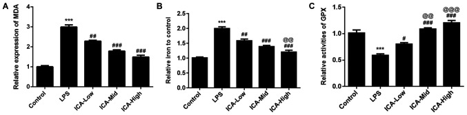
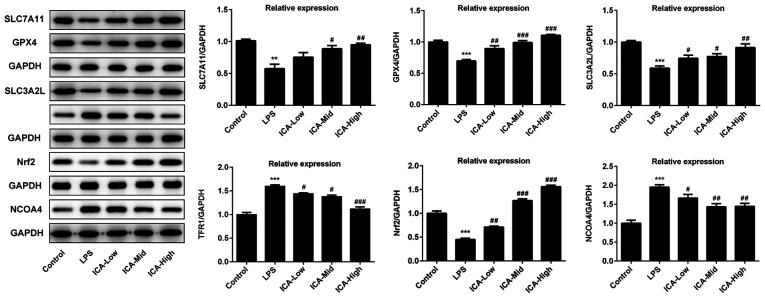
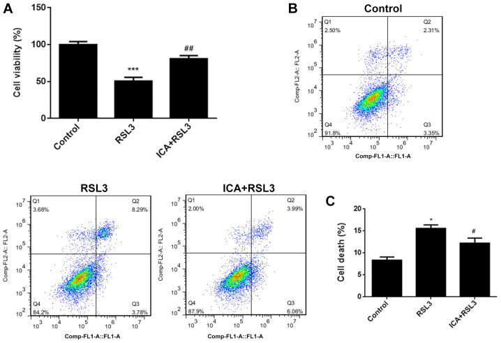
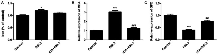
The mechanism of action of synovitis, as the vital pathological process of rheumatoid arthritis and osteoarthritis, remains to be elucidated. The effects and the mechanism of icariin (ICA), which is a promising therapeutic agent in synovitis, was investigated in the present study. In addition, ferroptosis, a vital cell process involved in several diseases, was also studied in synovitis for the first time. Lipopolysaccharide (LPS)-induced synoviocytes served as a synovitis cell model. The cells were divided into control, LPS and experimental groups and were treated with different concentrations of ICA. Cell viability was determined by Cell Counting Kit-8 assay and cell death was determined by flow cytometry. The expression levels of proteins (GPX4, SLC7A11, SLC3A2L, TRF, Nrf2 and NCOA4) were measured by western blotting. Quantification of malondialdehyde (MDA), iron and glutathione peroxidase 4 (GPX4) activity levels were performed via using corresponding assay kits. Cell death was increased, and cell viability was decreased in LPS-induced synoviocytes. Furthermore, MDA levels and iron content were elevated and GPX levels was reduced in LPS-induced synoviocytes. Transferrin receptor protein 1 and nuclear receptor coactivator 4 were upregulated and proteins of the Xc-/GPX4 axis, as well as nuclear factor erythroid 2-related factor 2, were decreased by LPS treatment. All aforementioned LPS affects were alleviated by ICA via a concentration-dependent manner. ICA counteracted the effects of RSL3, a ferroptosis activator, on cell viability, lipid peroxidation, iron content and relative protein expression of ferroptosis in synoviocytes. ICA protects the cells from death in synoviocytes induced by LPS, via the inhibition of ferroptosis by activating the Xc-/GPX4 axis, which can be exploited as a new therapeutic strategy for synovitis.

Keywords: Xc-/glutathione peroxidase 4 axis; ferroptosis; icariin; synoviocytes; synovitis.


Figures

Figure 1
Figure 1
The effects of ICA on cell viability and death. (A) Cell viability and (B and C) cell death in the different study groups. Experimental data were obtained from five independent experiments and shown as the mean ± standard deviation. ***P<0.001 vs. control; #P<0.05, ##P<0.01 and ###P<0.001 vs. LPS; @P<0.05 vs. ICA-Low; &&P<0.01 vs. ICA-Mid. ICA, icariin; ICA-Low, 2 µM ICA; ICA-Mid, 5 µM ICA; ICA-High, 10 µM ICA; LPS, lipopolysaccharide.
Figure 2
Figure 2
Effects of ICA on the levels of MDA, iron content and GPX. The levels of (A) MDA, (B) iron content and (C) GPX in the study groups. Experimental data were obtained from five independent experiments and shown as the mean ± standard deviation. @@P<0.01 and @@@P<0.001 vs. ICA-Low; ***P<0.001 vs. control; #P<0.05, ##P<0.01 and ###P<0.001 vs. LPS. ICA, icariin; ICA-Low, 2 µM ICA; ICA-Mid, 5 µM ICA; ICA-High, 10 µM ICA; LPS, lipopolysaccharide; MDA, malondialdehyde; GPX, glutathione peroxidase.
Figure 3
Figure 3
Effects of ICA on the levels of ferroptosis-associated proteins. The levels of proteins associated with ferroptosis in the study groups. The representative blot of five independent experiments with similar results are expressed as the mean ± standard deviation. ***P<0.001 vs. control; **P<0.01 vs. control; #P<0.05, ##P<0.01 and ###P<0.001 vs. LPS. ICA, icariin; ICA-Low, 2 µM ICA; ICA-Mid, 5 µM ICA; ICA-High, 10 µM ICA; LPS, lipopolysaccharide; SLC7A111, cystine/glutamate transporter; GPX, glutathione peroxidase; SLC3A2L, 4F2 cell-surface antigen heavy chain; TFR1, transferrin receptor protein 1; Nrf2, nuclear factor erythroid 2-related factor 2; NCOA4, nuclear receptor coactivator 4.
Figure 4
Figure 4
Effects of ICA on cell death and viability following inhibition of the Xc-/glutathione peroxidase 4 axis. (A) cell viability and (B and C) cell death in the different study groups. Experimental data were obtained from five independent experiments and shown as the mean ± standard deviation. *P<0.01 and ***P<0.001 vs. control; #P<0.05 and ##P<0.01 vs. RSL3. ICA, icariin.
Figure 5
Figure 5
Effects of ICA on the levels of MDA, iron content and GPX following inhibition of the Xc-/GPX4 axis. The levels of (A) MDA, (B) iron content and (C) GPX in the study groups. Bar graphs were drawn from five independent experiments. Results were expressed as the mean ± standard deviation. *P<0.05 and ***P<0.001 vs. control; ##P<0.01 and ###P<0.001 vs. RSL3. ICA, icariin; MDA, malondialdehyde; GPX, glutathione peroxidase.
Figure 6
Figure 6
Effects of ICA on ferroptosis-associated protein expression following inhibition of the Xc-/GPX4 axis. The expression levels of ferroptosis-associated proteins in the study groups. The protein levels been standardized against GAPDH expression. Values are expressed as the mean ± standard deviation. **P<0.01 and ***P<0.001 vs. control; #P<0.05, ##P<0.01 and ###P<0.001 vs. RSL3. ICA, icariin; SLC7A111, cystine/glutamate transporter; GPX, glutathione peroxidase; SLC3A2L, 4F2 cell-surface antigen heavy chain; TFR1, transferrin receptor protein 1; Nrf2, nuclear factor erythroid 2-related factor 2; NCOA4, nuclear receptor coactivator 4.

Similar articles

Cited by

References

    1. Smolen JS, Aletaha D, McInnes IB. Rheumatoid arthritis. Lancet. 2016;388:2023–2038. doi: 10.1016/S0140-6736(16)30173-8. - DOI - PubMed
    1. Safiri S, Kolahi AA, Hoy D, Smith E, Bettampadi D, Mansournia MA, Almasi-Hashiani A, Ashrafi-Asgarabad A, Moradi-Lakeh M, Qorbani M, et al. Global, regional and national burden of rheumatoid arthritis 1990-2017: A systematic analysis of the global burden of disease study 2017. Ann Rheum Dis. 2019;78:1463–1471. doi: 10.1136/annrheumdis-2019-215920. - DOI - PubMed
    1. Smolen JS, Aletaha D, Barton A, Burmester GR, Emery P, Firestein GS, Kavanaugh A, McInnes IB, Solomon DH, Strand V, Yamamoto K. Rheumatoid arthritis. Nat Rev Dis Primers. 2018;4(18001) doi: 10.1038/nrdp.2018.1. - DOI - PubMed
    1. Orsolini G, Fassio A, Rossini M, Adami G, Giollo A, Caimmi C, Idolazzi L, Viapiana O, Gatti D. Effects of biological and targeted synthetic DMARDs on bone loss in rheumatoid arthritis. Pharmacol Res. 2019;147(104354) doi: 10.1016/j.phrs.2019.104354. - DOI - PubMed
    1. Lazzerini PE, Capecchi PL, Laghi-Pasini F. Systemic inflammation and arrhythmic risk: Lessons from rheumatoid arthritis. Eur Heart J. 2017;38:1717–1727. doi: 10.1093/eurheartj/ehw208. - DOI - PubMed