

Posttraumatic synchronous double acute epidural hematomas: Two craniotomies, single skin incision
- PMID: 33365197
- PMCID: PMC7749931
- DOI: 10.25259/SNI_697_2020
Posttraumatic synchronous double acute epidural hematomas: Two craniotomies, single skin incision
Abstract
Background: Double epidural hematomas (EDHs) have a higher mortality rate compared to single EDHs and same Glasgow Coma Scale (GCS), although double EDHs incidence is less common.
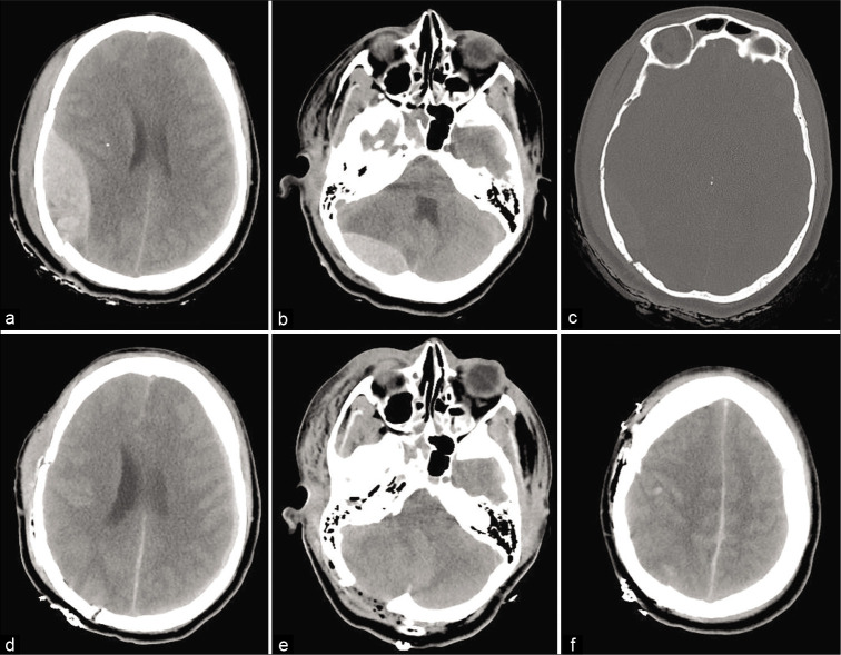
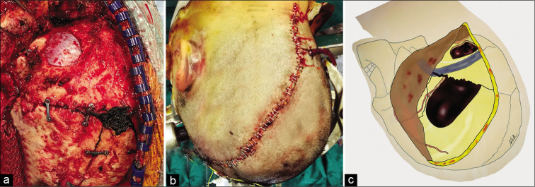
Case description: We present the case of a 34-year-old female who underwent single skin incision and frontotemporal and suboccipital craniotomies for fatal traumatic double acute EDHs, then, a literature review was performed.
Conclusion: Double EDHs in association with low GCS at presentation and traumatic diastasis of cranial sutures or other maxillofacial injuries are associated to an unfavorable outcome.
Keywords: Epidural hematoma; Neurotrauma; Traumatic brain injury.
Copyright: © 2020 Surgical Neurology International.
Conflict of interest statement
There are no conflicts of interest.
Figures
References
-
- Benedetto N, Gambacciani C, Montemurro N, Morganti R, Perrini P. Surgical management of acute subdural haematomas in elderly: Report of a single center experience. Br J Neurosurg. 2017;31:244–8. - PubMed
-
- Bullock MR, Chesnut R, Ghajar J, Gordon D, Hartl R, Newell DW, et al. Surgical management of acute epidural hematomas. Neurosurgery. 2006;58(Suppl 3):S7–15. - PubMed
-
- Fricia M, Umana GE, Scalia G, Raudino G, Passanisi M, Spitaleri A, et al. Posttraumatic triple acute epidural hematomas: First report of bilateral synchronous epidural hematoma and a third delayed. World Neurosurg. 2020;133:212–5. - PubMed
Publication types
LinkOut - more resources
Full Text Sources