. 2020 Dec 7:10:607253.
doi: 10.3389/fcimb.2020.607253. eCollection 2020.

Study on the Occurrence of Genetic Exchange Among Parasites of the Leishmania mexicana Complex

Affiliations

Study on the Occurrence of Genetic Exchange Among Parasites of the Leishmania mexicana Complex

Roman Telittchenko et al. Front Cell Infect Microbiol. .

Abstract

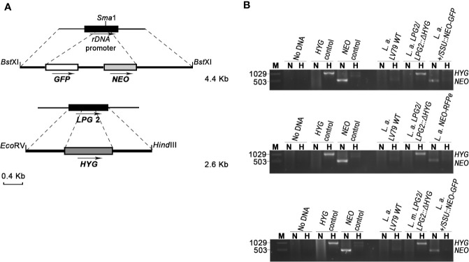
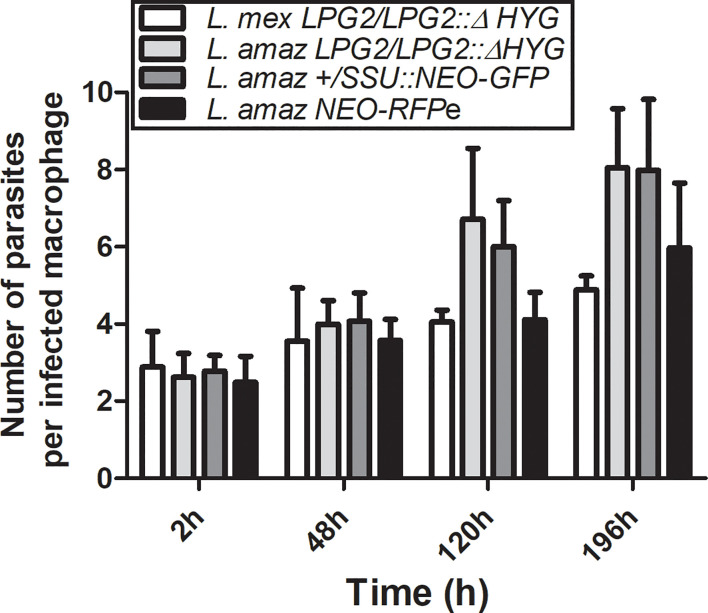
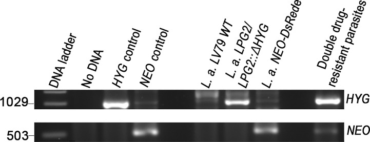
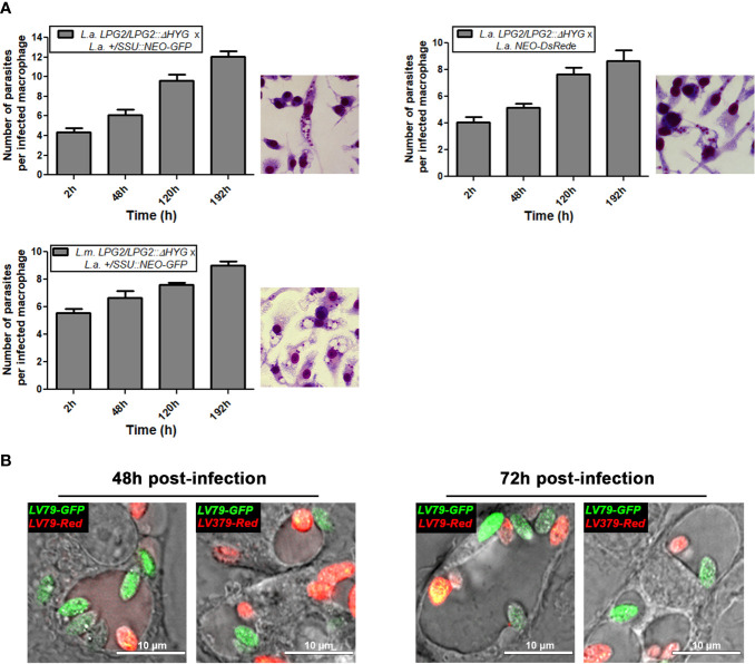
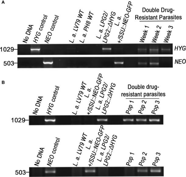
In Leishmania, genetic exchange has been experimentally demonstrated to occur in the sand fly vector and in promastigote axenic cultures through a meiotic-like process. No evidence of genetic exchange in mammalian hosts have been reported so far, possibly due to the fact that the Leishmania species used in previous studies replicate within individual parasitophorous vacuoles. In the present work, we explored the possibility that residing in communal vacuoles may provide conditions favorable for genetic exchange for L. mexicana and L. amazonensis. Using promastigote lines of both species harboring integrated or episomal drug-resistance markers, we assessed whether genetic exchange can occur in axenic cultures, in infected macrophages as well as in infected mice. We obtained evidence of genetic exchange for L. amazonensis in both axenic promastigote cultures and infected macrophages. However, the resulting products of those putative genetic events were unstable as they did not sustain growth in subsequent sub-cultures, precluding further characterization.

Keywords: Leishmania; drug resistance; genetic exchange; host-pathogen relationship; intracellular pathogen; macrophage.


Conflict of interest statement

The authors declare that the research was conducted in the absence of any commercial or financial relationships that could be construed as a potential conflict of interest.

Figures

Figure 1
Figure 1
Generation of drug-resistant Leishmania parasites. (A) L.d-rDNA-NEO-GFP and LPG2::ΔHYG targeting constructs were used for the integration into the ribosomal RNA locus or in one allele of LPG2, respectively. For the L.d-rDNA-NEO-GFP construct, the NEO-GFP resistance cassette (white and gray boxes) was inserted in the SmaI site of the ribosomal RNA locus (black rectangle). The dashed lines delimit the regions of recombination between the target genes and targeting constructs. Arrows indicate orientation. (B) PCR products for drug resistance markers HYG and NEO of L. amazonensis and L. mexicana parental strains. The size of HYG and NEO resistance genes is 1,029 and 503 bp long, respectively. The pLeish-HYG-GFP and the pKS-NEO-DsRed constructs were used as controls for the HYG and NEO genes, respectively. L.a. LV79 WT is a DNA sample used to show that our wild type parasites do not harbor any drug-resistance markers in their genomes. L. amazonensis LPG2/LPG2::ΔHYG, L. amazonensis +/SSU::NEO-GFP, L. mexicana LPG2/LPG2::ΔHYG and L. amazonensis NEO-DsRede are controls used to validate the presence of HYG and NEO resistance genes. No DNA sample was loaded as negative control. M, molecular DNA ladder; H, Hygromycin; N, G418.
Figure 2
Figure 2
Survival of parental strains within infected macrophages. BMMs were infected with metacyclic serum-opsonized promastigotes of L. amazonensis and L. mexicana parental strains (L. amazonensis LPG2/LPG2::ΔHYG, L. mexicana LPG2/LPG2::ΔHYG, L. amazonensis +/SSU::NEO-GFP, L. amazonensis NEO-DsRede) for 2, 48, 120, and 196 h. Bars represent mean ± SE of three representative experiments performed in triplicate in bone marrow derived murine macrophages. Parasites were counted in 100 macrophages and quantified by light microscopy.
Figure 3
Figure 3
Molecular genotype characterization of double drug-resistant parasites isolated from axenic cultures. PCR amplification of genes encoding antibiotic resistance. The size of HYG and NEO resistance genes is 1,029 and 503 bp long. The pLeish-HYG-GFP and the pKS-NEO-DsRed constructs were used as controls for the HYG and NEO genes, respectively. L.a. LV79 WT is a DNA sample used to show that our wild type parasites do not express any drug-resistance markers. L. amazonensis LPG2/LPG2::ΔHYG and L. amazonensis NEO-DsRede are controls used to validate the presence of HYG and NEO resistance genes within the appropriate parental strains. No DNA sample was loaded as negative control.
Figure 4
Figure 4
Survival of mating crosses within infected macrophages and visualization of both parental strains within the same vacuole. (A) BMMs were infected with metacyclic serum-opsonized promastigote crosses of L. mexicana complex parental parasite strains (L. amazonensis LPG2/LPG2::ΔHYG + L. amazonensis +/SSU::NEO-GFP; L. amazonensis LPG2/LPG2::ΔHYG + L. amazonensis NEO-DsRede; L. mexicana LPG2/LPG2::ΔHYG + L. amazonensis +/SSU::NEO-GFP for 2, 48, 120, and 196 h. Bars represent mean ± SE of three representative experiments performed in triplicate in bone marrow derived murine macrophages. Parasites were counted in 100 macrophages and quantified by light microscopy. Macrophages were stained with HEMA 3 kit. Representative pictures from each cross are shown. (B) Live microscopy analysis of L. amazonensis and L. mexicana parasite strains expressing different fluorescent markers. Representative pictures of both parental strains within the same communal vacuole at 48 and 72 h are shown. LV79-GFP, L. amazonensis HYG-GFPe; LV79-DsRed, L. amazonensis NEO-DsRede; M379-DsRed, L. mexicana NEO-DsRede.
Figure 5
Figure 5
Molecular genotype characterization of double drug-resistant parasites isolated from in vitro infections. PCR amplification of genes encoding antibiotic resistance. The size of HYG and NEO resistance genes is 1,029 and 503 bp long. The pLeish-HYG-GFP and the pKS-NEO-DsRed constructs were used as controls for the HYG and NEO genes, respectively. L.a. LV79 WT and L.a. PH8 WT is a DNA sample used to show that our wild type parasites do not express any drug-resistance markers. L. amazonensis LPG2/LPG2::ΔHYG, L. amazonensis +/SSU::NEO-GFP are controls used to validate the expression of HYG and NEO resistance genes within the appropriate parental strains. No DNA sample was loaded as negative control. (A) PCR amplification of resistance genes of the first double drug-resistant parasite population. Population was maintained for 3 weeks until it lost one of the resistance genes and perished. PCRs of double drug-resistant parasites represent the presence of both genes on weeks 1, 2, and 3 (B) PCR amplification of resistance genes of the second occurrence of double drug-resistant parasites. Three populations were isolated (Pop 1–3). Two of the population were found to be double-drug resistant and one was not.

References

    1. Akopyants N. S., Kimblin N., Secundino N., Patrick R., Peters N., Lawyer P., et al. (2009). Demonstration of genetic exchange during cyclical development of Leishmania in the sand fly vector. Science 324 (5924), 265–268. 10.1126/science.1169464 - DOI - PMC - PubMed
    1. Arango Duque G., Jardim A., Gagnon E., Fukuda M., Descoteaux A. (2019). The host cell secretory pathway mediates the export of Leishmania virulence factors out of the parasitophorous vacuole. PLoS Pathog. 15 (7), e1007982. 10.1371/journal.ppat.1007982 - DOI - PMC - PubMed
    1. Atayde V. D., da Silva Lira Filho A., Chaparro V., Zimmermann A., Martel C., Jaramillo M., et al. (2019). Exploitation of the Leishmania exosomal pathway by Leishmania RNA virus 1. Nat. Microbiol. 4 (4), 714–723. 10.1038/s41564-018-0352-y - DOI - PubMed
    1. Banuls A. L., Guerrini F., Le Pont F., Barrera C., Espinel I., Guderian R., et al. (1997). Evidence for hybridization by multilocus enzyme electrophoresis and random amplified polymorphic DNA between Leishmania braziliensis and Leishmania panamensis/guyanensis in Ecuador. J. Eukaryot. Microbiol. 44 (5), 408–411. 10.1111/j.1550-7408.1997.tb05716.x - DOI - PubMed
    1. Banuls A. L., Jonquieres R., Guerrini F., Le Pont F., Barrera C., Espinel I., et al. (1999). Genetic analysis of leishmania parasites in Ecuador: are Leishmania (Viannia) panamensis and Leishmania (V.) Guyanensis distinct taxa? Am. J. Trop. Med. Hyg. 61 (5), 838–845. 10.4269/ajtmh.1999.61.838 - DOI - PubMed

Publication types

Grants and funding