. 2020 Dec 16:25:100871.
doi: 10.1016/j.bbrep.2020.100871. eCollection 2021 Mar.

Functional expression of human prostaglandin E2 receptor 4 (EP4) in E. coli and characterization of the binding property of EP4 with Gα proteins

Affiliations

Functional expression of human prostaglandin E2 receptor 4 (EP4) in E. coli and characterization of the binding property of EP4 with Gα proteins

Nam Hyuk Kim et al. Biochem Biophys Rep. .

Abstract

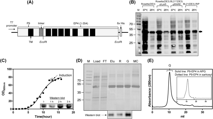
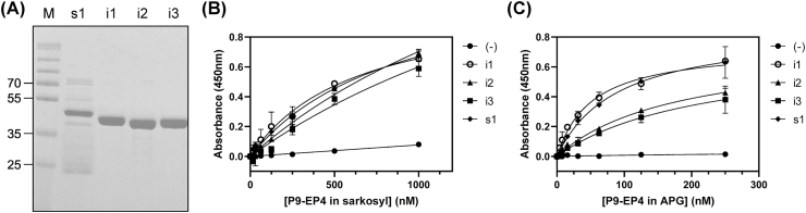
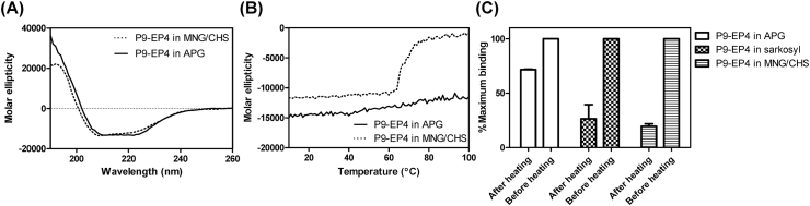
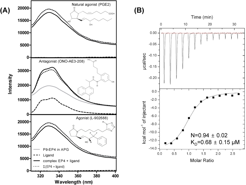
Human prostaglandin E2 receptor 4 (EP4) is one of the four subtypes of prostaglandin E2 (PGE2) receptors and belongs to the rhodopsin-type G protein-coupled receptor (GPCR) family. Particularly, EP4 is expressed in various cancer cells and is involved in cancer-cell proliferation by a G protein signaling cascade. To prepare an active form of EP4 for biochemical characterization and pharmaceutical application, this study designed a recombinant protein comprising human EP4 fused to the P9 protein (a major envelope protein of phi6 phage) and overexpressed the P9-EP4 fusion protein in the membrane fraction of E. coli. The solubilized P9-EP4 with sarkosyl (a strong anionic detergent) was purified by affinity chromatography. The purified protein was stabilized with amphiphilic polymers derived from poly-γ-glutamate. The polymer-stabilized P9-EP4 showed specific interaction with the alpha subunits of Gs or Gi proteins, and a high content of α-helical structure by a circular dichroism spectroscopy. Furthermore, the polymer-stabilized P9-EP4 showed strong heat resistance compared with P9-EP4 in detergents. The functional preparation of EP4 and its stabilization with amphiphilic polymers could facilitate both the biochemical characterization and pharmacological applications targeting EP4.

Keywords: EP4; G protein; GPCR; Overexpression; PGE2; Purification.


Figures

Fig. 1
Fig. 1
Expression and purification of P9-EP4. (A) Schematic representation of the P9-EP4 expression vector. The coding sequence of human EP4 (amino acids 1–354) was linked to the C-terminus of the P9 protein. The transmembrane regions and the C-terminus His6-tag sequence are indicated as a black box and dotted box, respectively. (B) Expression test of P9-EP4 in various E. coli expression hosts. Cells from the culture after induction for 2 h at 37 °C (37 °C), or induction for 3 h at 25 °C (25 °C) were collected and resuspended in an SDS-PAGE sample buffer. The separated proteins were analyzed using western blotting with anti-P9 antibodies. The P9-EP4 band is indicated by an arrow. (C) Culture of BL21(DE3)-RIP cells harboring pP9-EP4 using a fermenter. The expression of P9-EP4 was induced by adding 1 mM IPTG when the OD600 nm of the culture media at 37 °C reached 70. Expression level of P9-EP4 in the cells before (−) and after 1, 2, and 3 h of induction was measured using western blotting with anti-P9 antibodies (inserted panel). (D) Purification of P9-EP4. The samples from each purification step were analyzed by SDS-PAGE and proteins were visualized using Coomassie staining. The lanes are described as follows. M: size marker, Load: soluble membrane fraction after solubilization with 1% sarkosyl, FT: flow-through of Ni-NTA chromatography, Elu: eluate from Ni-NTA chromatography, R: reconstituted P9-EP4 with APG, G: purified P9-EP4 stabilized with APG by gel filtration chromatography, and MC: purified EP4 in 0.05% MNG/0.005% CHS. Identical samples used for SDS-PAGE were analyzed using western blotting with anti-6X His-tag antibodies. (E) The elution chromatogram of gel filtration chromatography using Superdex200 10/300 GL of P9-EP4 stabilized with APG. The elution volume that contains P9-EP4 is indicated as G.
Fig. 2
Fig. 2
Measurement of the interaction between P9-EP4 and various G protein alpha subunits by ELISA. (A) The purified Gαi1 (i1), Gαi2 (i2), Gαi3 (i3), and Gαs1 (s1) were analyzed by SDS-PAGE followed by Coomassie staining. (B, C) The concentration-dependent binding of P9-EP4 in sarkosyl (B) or in APG (C) to the immobilized Gα proteins. Five to 1000 nM of P9-EP4 was applied to the plate treated with 5% of skimmed milk blocking solution (filled circle), or to the plate immobilized with 10 μg/ml of Gαi1 (open circle), Gαi2 (filled triangle), Gαi3 (filled square), or Gαs1 (filled diamond), and the amount of bound P9-EP4 was measured using an anti-P9 antibody.
Fig. 3
Fig. 3
Analysis of secondary structure and thermostability of P9-EP4. (A) The CD spectra of P9-EP4 stabilized with a mixture of MNG and CHS (0.1 mg/ml of P9-EP4 in 10 mM sodium phosphate pH 7.4, 0.05% MNG, and 0.005% CHS; dotted line) or P9-EP4 stabilized with APG (0.063 mg/ml of P9-EP4 in 10 mM sodium phosphate pH 7.4, 0.05% APG: solid line) in the wavelength of 190–260 nm. (B) Temperature-dependent change of the molar ellipticity of P9-EP4 in MNG/CHS (dotted line) or in APG (solid line) at the fixed wavelength of 220 nm. (C) Comparison of the residual binding activity of P9-EP4 to the immobilized Gαi1 after heat treatment. P9-EP4 in sarkosyl, MNG/CHS or APG were incubated at 80 °C for 30 min, and the amount of bound P9-EP4 was measured using anti-P9 antibodies and compared with the value of P9-EP4 before heat treatment.
Fig. 4
Fig. 4
Agonist- and antagonist-binding analysis by the intrinsic fluorescence spectrum of EP4 and ITC.(A) The effects of the endogenous ligand (PGE2), antagonist (ONO-AE3-208), and agonist (L-902688) on the intrinsic fluorescence spectrum of P9-EP4 in APG. The Trp fluorescence spectra of P9-EP4 in APG was measured at 310–400 nm using an excitation wavelength of 295 nm in the absence (gray lines) or presence (solid lines) of chemicals. The spectra of chemicals are indicated as broken lines, and the mathematical summed spectra of chemicals and P9-EP4 are indicated as dotted lines. (B) ITC analysis shows that ONO-AE3-208 binds EP4 with KD = 0.68 ± 0.15 μM at 1:1 M ratio. Raw ITC data (Top panel) and integrated heats of injection (bottom panel) are presented for the titration between EP4 and ONO-AE3-208. In the bottom panel, the experimental data are shown as solid squares and the least squares best-fit curves derived from a simple one-site binding model are shown as a black line.

Similar articles

Cited by

References

    1. Smith W.L., DeWitt D.L., Garavito R.M. Cyclooxygenases: structural, cellular, and molecular biology. Annu. Rev. Biochem. 2000;69:145–182. - PubMed
    1. Smith W.L., Marnett L.J., DeWitt D.L. Prostaglandin and thromboxane biosynthesis. Pharmacol. Ther. 1991;49:153–179. - PubMed
    1. Hara S., Kamei D., Sasaki Y., Tanemoto A., Nakatani Y., Murakami M. Prostaglandin E synthases: understanding their pathophysiological roles through mouse genetic models. Biochimie. 2010;92:651–659. - PubMed
    1. Montrose D.C., Nakanishi M., Murphy R.C., Zarini S., McAleer J.P., Vella A.T., Rosenberg D.W. The role of PGE2 in intestinal inflammation and tumorigenesis. Prostag. Other Lipid Mediat. 2015;116–117:26–36. - PMC - PubMed
    1. Murakami M., Naraba H., Tanioka T., Semmyo N., Nakatani Y., Kojima F., Ikeda T., Fueki M., Ueno A., Oh-ishi S. Regulation of prostaglandin E2 biosynthesis by inducible membrane-associated prostaglandin E2 synthase that acts in concert with cyclooxygenase-2. J. Biol. Chem. 2000;275:32783–32792. - PubMed

LinkOut - more resources