

Imagery - ultrasound biomicroscopy and anterior segment Optical Coherence Tomography - in the diagnosis of anterior segment pathology
- PMID: 33367163
- PMCID: PMC7739559
Imagery - ultrasound biomicroscopy and anterior segment Optical Coherence Tomography - in the diagnosis of anterior segment pathology
Abstract
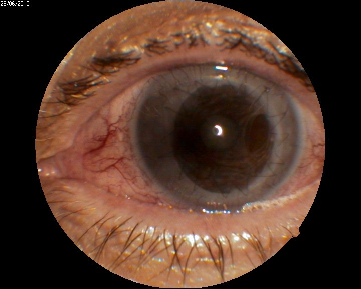
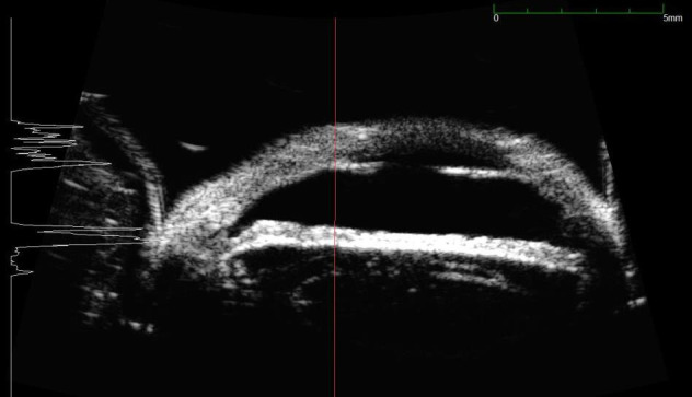
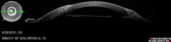
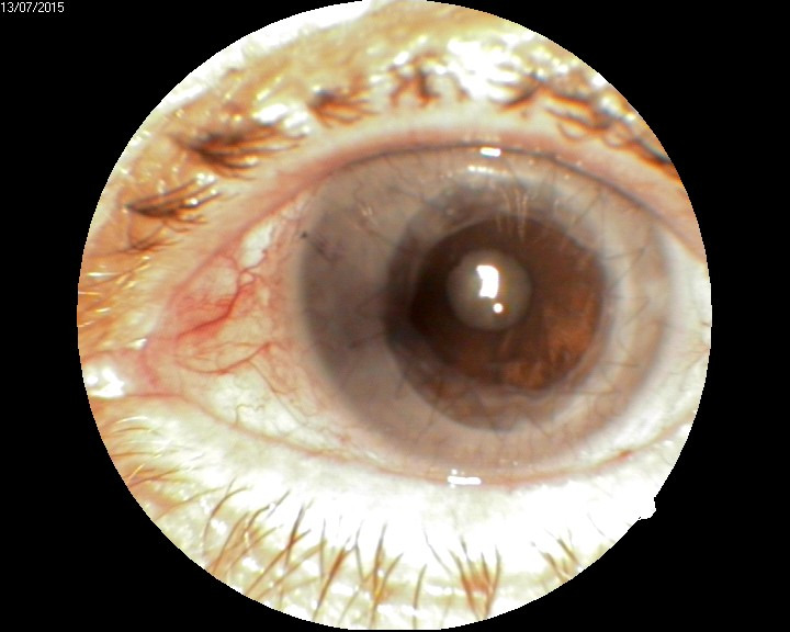
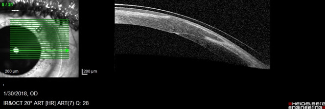
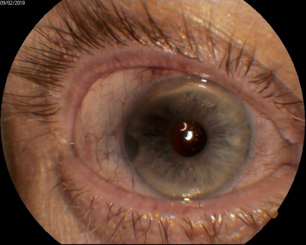
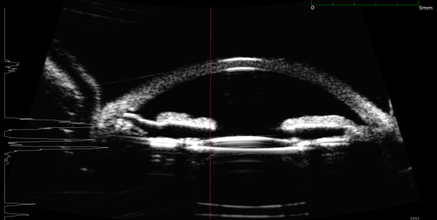
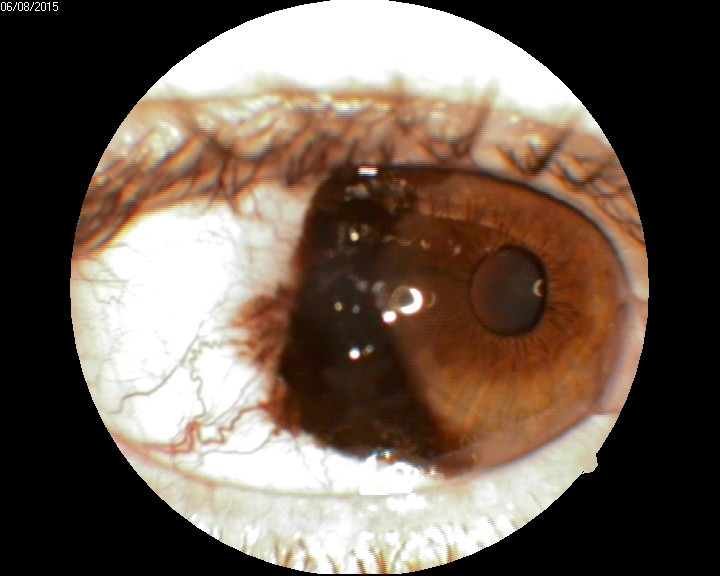
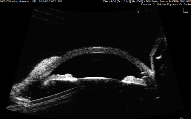
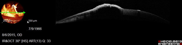
Purpose: The aim of this paper was to show the usefulness of imagery in better documenting the pathology of the anterior segment. Methods: The article comprises clinical cases, insisting on how imagery was essential in establishing the diagnosis or the therapeutic plan. Results: Lack of imagery would have made establishing a proper diagnosis much more difficult. Conclusions: Although clinical examination is simple and offers a fairly good amount of information, some particular cases of anterior segment pathology need additional investigations, every method having its indications and limits.
Keywords: anterior segment optical coherence tomography; ultrasound biomicroscopy.
©Romanian Society of Ophthalmology.
Figures
References
-
- Pavlin CJ, Harsiewicz K, Sherar MD, et al. Clinical use of ultrasound biomicroscopy. Ophthalmology. 1991;98(30):287–295. - PubMed
-
- Pavlin CJ, Foster FS. Ultrasound biomicroscopy of the Eye. New York: Springer Verlag; 1994.
-
- Woo EK, Pavlin CJ, Slomovic A, et al. Ultrasound biomicroscopic quantitative analysis of light-dark changes associated with pupillary block. Am J Ophthalmol. 1999;127:43–47. - PubMed
-
- Shukle S, Danmgi KF, Harasynowycz P, et al. Clinical features distinguishing angle-closure from pseudoplateau versus plateau iris. Br. J. Ophthalmol. 2008;92:340–344. - PubMed
-
- Bernstein DM, Gentile RC, Sidoti PA, et al. Ultrasound biomicroscopy in anterior ocular trauma. Ophthalmic Surg Lasers. 1997;28:201–207. - PubMed
Publication types
MeSH terms
LinkOut - more resources
Full Text Sources
Medical