Review
. 2020 Oct-Dec;64(4):323-332.
doi: 10.22336/rjo.2020.54.

Vascular emergencies in neuro-ophthalmology

Affiliations
Review

Vascular emergencies in neuro-ophthalmology

Eugenia Raluca Iorga et al. Rom J Ophthalmol. 2020 Oct-Dec.

Abstract

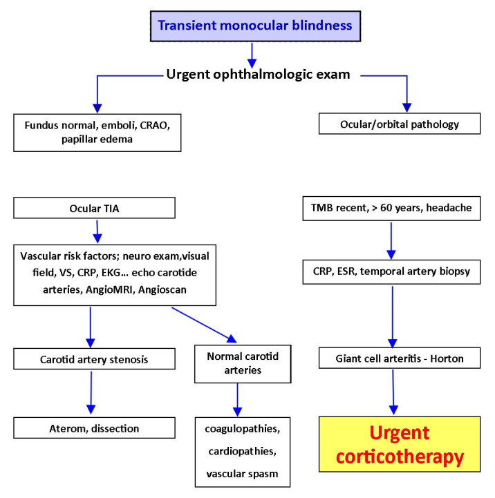
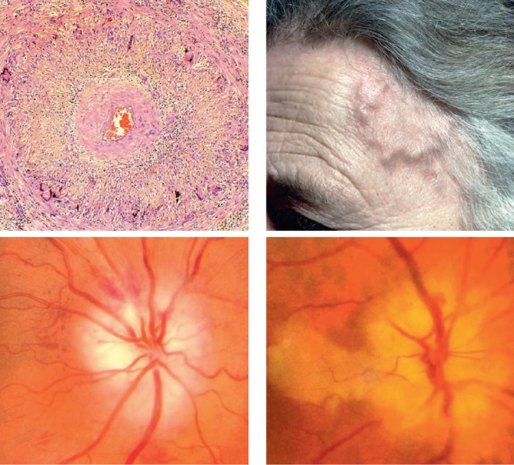
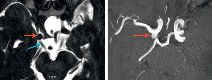
The cerebral vascularization is assured by the 2 internal carotids and 2 vertebral arteries, and the Willis circle. Carotid artery obstruction is the most common abnormality associated with ocular ischemic syndrome. Obstruction may be due to atheromatous plaque, external compression, arteritis, or dissection of the artery. An atheromatous lesion of the carotid artery is the most frequent lesion responsible for ocular ischemic syndrome. The signs and symptoms of ocular ischemic syndrome are associated with severe hypoperfusion of the eye. Inflammatory lesions of the carotid artery are responsible for decreased flow in the carotid system. Other vascular emergencies are carotid artery dissection, Horton arteritis, aneurysms and carotid-cavernous fistula. The most common ocular signs and symptoms are transient monocular blindness, persistent monocular blindness, ocular ischemia, Claude Bernard Horner syndrome and oculomotor palsies. The carotid pathology can be a life-threatening pathology and it is important to recognize all these signs and symptoms. A multi-specialty approach will prevent misdiagnosis and lead to a better patient management. Abbreviations: OIS = ocular ischemic syndrome, TMB = transient monocular blindness, TIA = transient ischemic attack, ESR = erythrocyte sedimentation rate, CRP = C reactive protein, NVE = neovascularization elsewhere in the retina, NVD = neovascularization on the disc, AION A = anterior ischemic arteritic optic neuropathy, CBH = Claude Bernard Horner syndrome, MRI = magnetic resonance imaging.

Keywords: Claude Bernard Horner syndrome; carotid artery stenosis; ocular ischemic syndrome; oculomotor palsies; transient monocular blindness.


Figures

Fig. 1
Fig. 1
Cerebral and ocular vascularization. a. Cerebral vascularization. 1: anterior cerebral artery; 2: middle cerebral artery; 3: anterior communicating artery; 4: artery internal carotid; 5: common carotid artery; 6: superficial temporal artery; 7: ophthalmic artery; 8: supraorbital artery; 9: artery supratrochlear; 10: median palpebral artery; 11: dorsal artery of the nose; 12: lateral palpebral artery; 13: angular artery; 14: artery lacrimal; 15: transverse facial artery; 16: maxillary artery; 17: middle meningeal artery; 18: facial artery; 19: external carotid artery. b. Vascularization of the eyeballs by ophthalmic artery, which is a branch of the internal carotid artery. (from Vignal-Clermont C, Tilikete C, Milea D. Neuro-ophtalmologie. 2e edition, 2016, Elsevier)
Fig. 2
Fig. 2
Angio-scanner (a) and MRA (b) of the supra-aortic trunks, carotid Doppler (c). Tight right carotid stenosis (from Vignal-Clermont C, Tilikete C, Milea D. Neuro-ophtalmologie. 2e edition, 2016, Elsevier)
Fig. 3
Fig. 3
Management of the transient monocular blindness
Fig. 4
Fig. 4
Giant cell arteritis - Horton. A) Histology - granulomatous inflammation and narrowing of the lumen; (B) the superficial temporal artery is pulseless and thickened; (C) pale swollen disc; (D) papillary oedema and cilioretinal artery occlusion (from Bowling B. Kanski’s Clinical Ophthalmology. A systematic approach. Eighth edition, 2016, Elsevier)
Fig. 5
Fig. 5
Angio MRI of the supra aortic trunks (a); MRI with diffusion slices B1000 (b); T1 gadolinium FATSAT (c,d); Carotid hematoma (c) with carotid stenosis (a). Carotid dissection with intracranial extension (d) (from Vignal-Clermont C, Tilikete C, Milea D. Neuro-ophtalmologie. 2e edition, 2016, Elsevier)
Fig. 6
Fig. 6
Carotid dissection - painful Claude Bernard Horner (from DU Neuro-ophtalmologie, 2018-2019, Paris, Pupilles, Lamirel)
Fig. 7
Fig. 7
Post-traumatic carotid-cavernous fistula - ptosis, exophthalmos and hypotropia of the eye right (a), global ocular motility disorders (b-d). The episcleral veins are evident (d). Angio-scanner - visualization of the carotid-cavernous fistula and dilation of the ophthalmic vein (e) (from Vignal-Clermont C, Tilikete C, Milea D. Neuro-ophtalmologie. 2e edition, 2016, Elsevier)
Fig. 8
Fig. 8
MRI axial T2 (a) and TOF (b). Posterior communicating artery aneurysm compressing the IIIrd oculomotor nerve (from Vignal-Clermont C, Tilikete C, Milea D. Neuro-ophtalmologie. 2e edition, 2016, Elsevier)
Fig. 9
Fig. 9
Complete III nerve palsy right eye. - complete right ptosis (a) the eye is in divergence and hypotropia, (b) pupil - mydriasis, (c) impossibility of elevation, (d) adduction and (e) lowering of the globe (from Vignal-Clermont C, Tilikete C, Milea D. Neuro-ophtalmologie. 2e edition, 2016, Elsevier)

References

    1. Rea P. Blood Supply of the Brain and Clinical Issues. Essential Clinical Anatomy of the Nervous System. 2015
    1. Ashrafian H. Anatomically specific clinical examination of the carotid arterial tree. Anatomical Science International. 2007 Mar;82(1):16–23. - PubMed
    1. Brown GC, Green WR. The ocular ischaemic syndrome. Curr Opinion Ophthalmol. 1994;5:14–20.
    1. Crassard I. Oeil. Pathologie carotid et vertebrobasilaire. DU Neuro-ophtalmologie. Paris: 2018-2019.
    1. Solomon CG, Grotta JC. Carotid Stenosis. New England Journal of Medicine. 2013;369(12):1143–1150. - PubMed