Toric intraocular lens implantation - atypical cases
- PMID: 33367183
- PMCID: PMC7739021
- DOI: 10.22336/rjo.2020.67
Toric intraocular lens implantation - atypical cases
Abstract
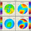
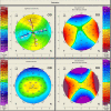
Objective: To describe the results of toric intraocular lens (IOL) implantation in three atypical cases (four eyes) with cataract and corneal astigmatism: one with bilateral keratoconus, one with pellucid marginal degeneration and one with buphthalmos due to congenital glaucoma. Methods: Three patients (four eyes) with corneal astigmatism (one with bilateral keratoconus, one with pellucid marginal degeneration and one with buphthalmos due to congenital glaucoma) underwent cataract surgery by standard phacoemulsification and the implantation of toric IOLs in the capsular bag. The presence of corneal astigmatism was identified by automated keratometry and confirmed by Scheimpflug-based corneal tomography. The toric IOL implanted in all cases was a single-piece AcrySof Toric IOL (Alcon Laboratories, Inc.). Postoperative visual acuity, the reduction in the refractive astigmatism, the spherical equivalent (SE) and the rotational stability of the toric IOL were recorded for all the patients. Results: Visual acuity increased and the refractive astigmatism decreased in all cases. In Case 1, the right eye achieved a postoperative uncorrected visual acuity (UCVA) of 20/ 20, a decrease in the refractive astigmatism from -3 DCyl to -0.75 DCyl and a spherical equivalent (SE) of -0.25. The left eye presented with a best-corrected visual acuity (BCVA) of 20/ 20, a decrease in the refractive astigmatism from -1.50 DCyl to -1.25 DCyl and a SE of -0.25. In Case 2, the postoperative UCVA was 20/ 20, with a decrease in the refractive astigmatism from -5.5 DCyl to -1 DCyl and a SE for the right eye of 0.00 D. In Case 3, the postoperative BCVA was 20/ 20, with a decrease in the refractive astigmatism from -4.75 DCyl to -1.50 DCyl and a SE of +1.25. No misalignment of the axis of the toric IOL was observed in any patient at subsequent follow-ups. The postoperative visual acuity was satisfactory for all the patients. Conclusions: Toric intraocular lenses can be an effective option for implantation in patients with cataract and corneal astigmatism in atypical situations such as mild to moderate keratoconus, pellucid marginal degeneration and buphthalmos due to congenital glaucoma. Predicting the refractive outcome is difficult in atypical cases and the surgeon should have accuracy and consistency in the preoperative measurements, for achieving satisfactory postoperative results.
Keywords: astigmatism; buphthalmos; cataract; congenital glaucoma; keratoconus; pellucid marginal degeneration; phacoemulsification; toric intraocular lens.
©Romanian Society of Ophthalmology.
Figures


References
-
- Shimizu K, Misawa A, Suzuki Y. Toric intraocular lenses: correcting astigmatism while controlling axis shift. J Cataract Refract Surg. 1994;20(5):523–526. - PubMed
-
- Nanavaty MA, Lake DB, Daya SM. Outcomes of pseudophakic toric intraocular lens implantation in Keratoconic eyes with cataract. J Refract Surg. 2012;28(12):884–889. - PubMed
-
- Luck J. Customized ultra-high-power toric intraocular lens implantation for pellucid marginal degeneration and cataract. J Cataract Refract Surg. 2010;36(7):1235–1238. - PubMed
-
- Stewart CM, McAlister JC. Comparison of grafted and non-grafted patients with corneal astigmatism undergoing cataract extraction with a toric intraocular lens implant. Clin Experiment Ophthalmol. 2010;38(8):747–757. - PubMed
-
- Alfonso JF, Fernández-Vega L, Lisa C, Fernandes P, González-Méijome JM, Montés-Micó R. Collagen copolymer toric posterior chamber phakic intraocular lens in eyes with keratoconus. Journal of Cataract & Refractive Surgery. 2010;36(6):906–916. - PubMed
Publication types
MeSH terms
LinkOut - more resources
Full Text Sources
Medical
