. 2021 Jun 15;9(12):4260-4277.
doi: 10.1039/d0bm01497d.

Milk exosomes with enhanced mucus penetrability for oral delivery of siRNA

Affiliations

Milk exosomes with enhanced mucus penetrability for oral delivery of siRNA

Matthew R Warren et al. Biomater Sci. .

Abstract

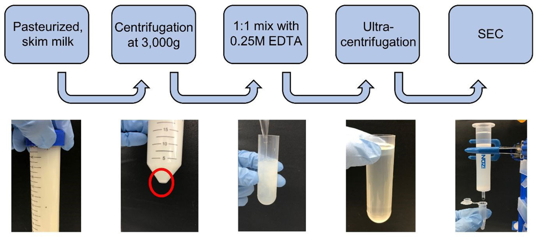
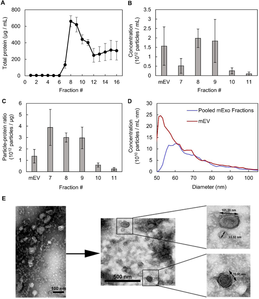
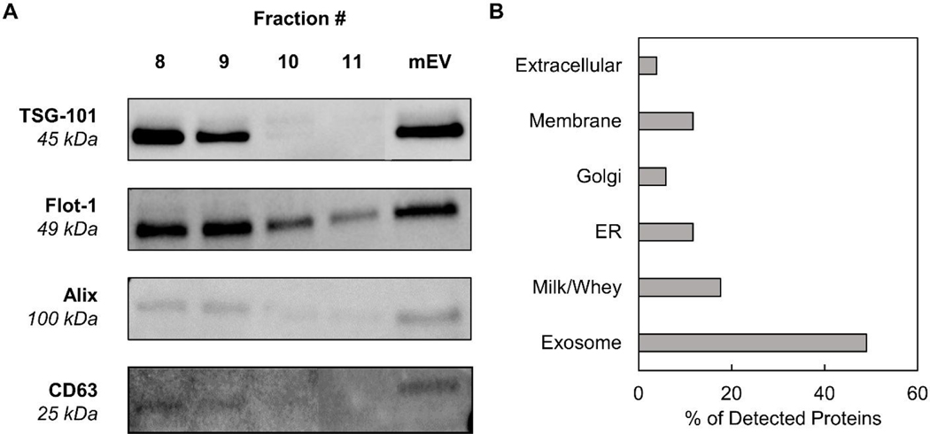
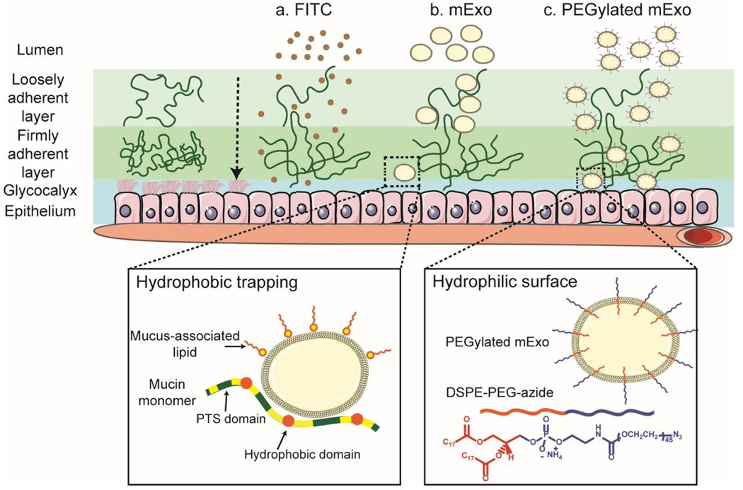
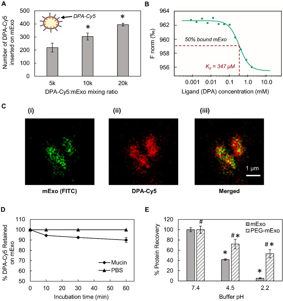
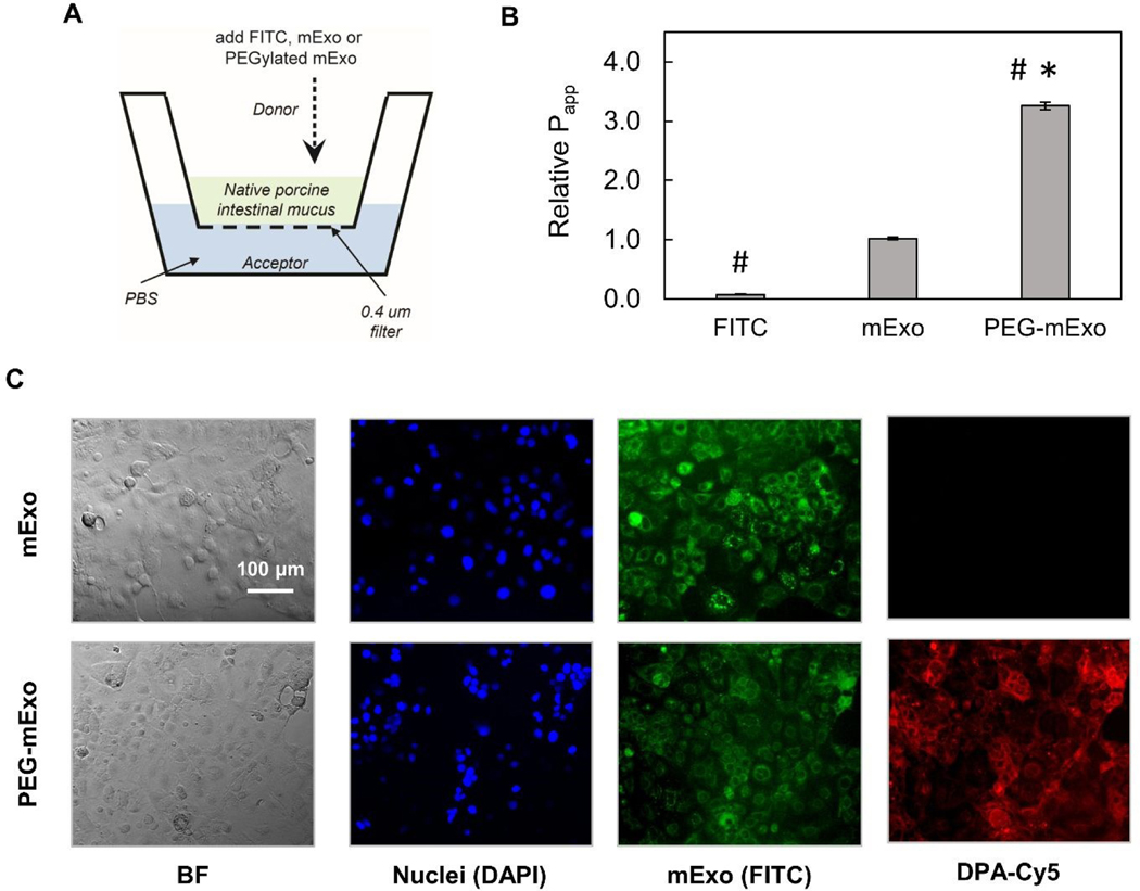
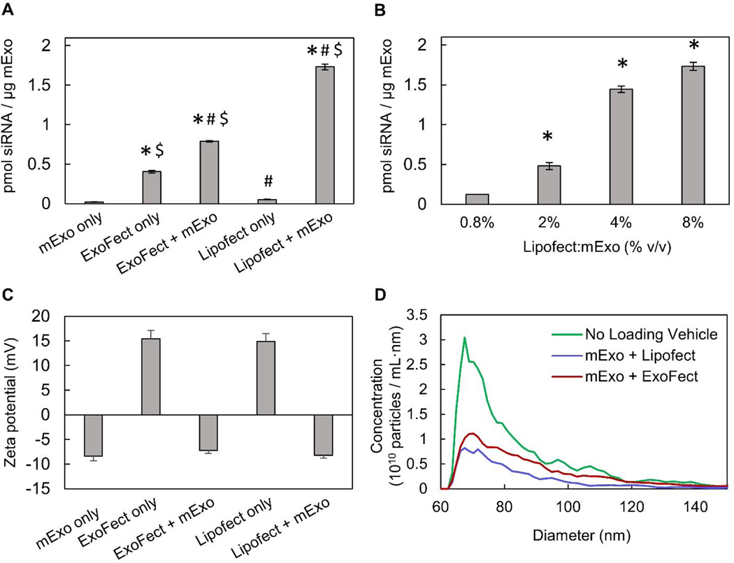
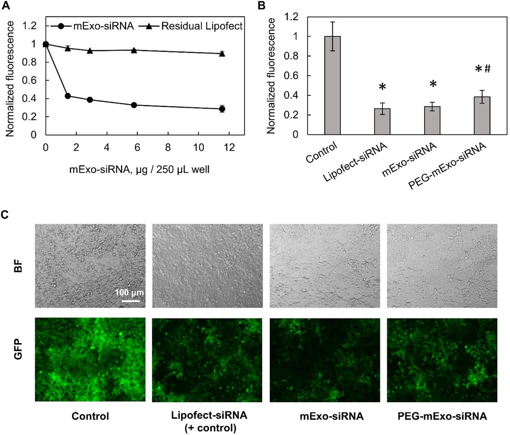
Bovine milk-derived exosomes have recently emerged as a promising nano-vehicle for the encapsulation and delivery of macromolecular biotherapeutics. Here we engineer high purity bovine milk exosomes (mExo) with modular surface tunability for oral delivery of small interfering RNA (siRNA). We utilize a low-cost enrichment method combining casein chelation with differential ultracentrifugation followed by size exclusion chromatography, yielding mExo of high concentration and purity. Using in vitro models, we demonstrate that negatively charged hydrophobic mExos can penetrate multiple biological barriers to oral drug delivery. A hydrophilic polyethylene glycol (PEG) coating was introduced on the mExo surface via passive, stable hydrophobic insertion of a conjugated lipid tail, which significantly reduced mExo degradation in acidic gastric environment and enhanced their permeability through mucin by over 3× compared to unmodified mExo. Both mExo and PEG-mExo exhibited high uptake by intestinal epithelial cells and mediated functional intracellular delivery of siRNA, thereby suppressing the expression of the target green fluorescence protein (GFP) gene by up to 70%. We also show that cationic chemical transfection is significantly more efficient in loading siRNA into mExo than electroporation. The simplicity of isolating high purity mExo in high concentrations and equipping them with tunable surface properties, demonstrated here, paves way for the development of mExo as an effective, scalable platform technology for oral drug delivery of siRNA.


Conflict of interest statement

Conflicts of Interest

There are no conflicts to declare.

Figures

Figure 1:
Figure 1:. Isolation and purification process of exosomes from bovine milk.
Pasteurized milk samples were first diluted with PBS and centrifuged at 3,000 g to pellet cells and debris. The supernatant was then treated with an equal volume of 0.25 M EDTA to chelate casein micelles and casein-coated exosomes. This was followed by differential ultracentrifugation to remove large protein aggregates and contaminating microvesicles. The sample was subsequently centrifuged at 100,000 g for 2 h to pellet extracellular vesicles (mEV). Finally, the resuspended mEV pellet was purified by size-exclusion chromatography (SEC) using a commercially available column.
Figure 2:
Figure 2:. Physical characteristics of individual mEV fractions following size exclusion chromatography.
(A) Total protein concentration of individual SEC fractions 1–16, measured by BCA assay. (B) Particle concentration in exosome fractions (7–11), measured by Spectradyne nCS1. (C) Purity of milk exosome (mExo) fractions, represented as particle-to-total protein ratio. (D) Representative size distribution of pre-SEC mEV sample and pooled fractions 8 and 9, measured by Spectradyne nCS1. (E) Transmission electron microscopy images of pooled mExo sample showing spherical morphology. Scale bars on left and middle images represent 100 nm and 500 nm, respectively. Data are presented as mean ± 95% confidence intervals (calculated by t-distribution).
Figure 3:
Figure 3:. Screening for the presence of protein exosome biomarkers in mExo samples.
(A) Western blotting for the presence of a variety of exosome markers, including membrane proteins CD63 and Flotillin-1 and ESCRT proteins (TSG-101 and Alix). Individual fractions after SEC are compared to the resuspended mEV pellet after ultracentrifugation and before SEC. (B) Breakdown of the categories of proteins present in exosome proteomics analysis by mass spectrometry.
Figure 4:
Figure 4:. Enhanced transport of mExo through intestinal mucus by surface PEGylation.
Intestinal mucus presents a significant barrier to oral drug delivery. Transport of unmodified mExo through the mucin is slowed down due to hydrophobic interactions between mExo lipid bilayer and hydrophobic domains of mucin. mExo was PEGylated by inserting the hydrophobic end of DSPE-PEG-azide (DPA) into its bilayer to enhance its effect on transport properties across intestinal mucus.
Figure 5:
Figure 5:. PEGylation of mExo and characterization.
(A) The number of DPA-Cy5 inserted on mExo surface for different mixing ratios (* vs 5k mixing ratio; p<0.05). (B) Binding affinity of DPA with the mExo bilayer using microscale thermophoresis. (C) Confocal microscopy images of dual-labeled PEG mExo. mExo is marked green; DPA is labeled red. Spatial overlap of green and red signal confirms insertion of DPA-Cy5 into the mExo membrane. (D) Stability of the DPA hydrophobic anchor on mExo surface over 1 h in the presence and absence of mucin. (E) Tolerability of unmodified and PEGylated mExo in conditions mimicking infant (pH = 4.5) and adult (pH = 2.2) stomach acidity. (* vs corresponding condition at pH 7.4, # vs mExo; p<0.05).
Figure 6:
Figure 6:. Transport of mExo and PEG-mExo through porcine intestinal mucus and uptake in human intestinal epithelial (Caco-2) cells.
(A) A transwell setup was used for evaluating FITC, mExo and PEG-mExo permeability in porcine intestinal mucin. (B) Relative permeability coefficients (Papp) of unmodified mExo and PEG-mExo in mucus. (* vs FITC, # vs mExo; p<0.05). (C) Uptake of mExo and PEG-mExo in Caco-2 cells after incubation for 2.5 h. mExo is labeled green, DPA in PEG-mExo is labeled red and cell nuclei are in blue. Control images accounting for background signal from free fluorophores are shown in Supplemental Figure S3.
Figure 7:
Figure 7:. Characterization of mExo-siRNA loaded via ExoFect and Lipofectamine
(A) Comparison of relative efficiencies of intra-luminal siRNA loading into mExo using two chemical transfection reagents, ExoFect and Lipofectamine-2000 (* vs Lipofect only, # vs ExoFect only, $ vs mExo only; p<0.05). (B) Optimization of mExo siRNA loading using Lipofect. (C) Change in particle surface charge following siRNA loading by ExoFect and Lipofectamine. (D) Change in particle size following loading by both methods. Data are presented as mean ± 95% bootstrap confidence intervals (* vs 0.8% Lipofect:mExo (% v/v); p<0.05).
Figure 8:
Figure 8:. mExo-mediated siRNA delivery and silencing of GFP in HEK293 cells.
(A) Dose-dependent response of exosome-mediated GFP silencing, compared to residual Lipofect-siRNA complexes left over from the purification process. (B) Comparison of silencing between unmodified and PEGylated mExo. (C) Fluorescence microscopy imaging showing silencing of GFP in confluent HEK293 cells. White scale bar represents 100 μm. (* vs Control, # vs mExo-siRNA; p<0.05).

Similar articles

Cited by

References

    1. Odom EBP, K.B.; Odom DC, Clinical Research, 2016, 8.
    1. Betker JL, Angle BM, Graner MW and Anchordoquy TJ, J Pharm Sci, 2019, 108, 1496–1505. - PMC - PubMed
    1. Shepard J, Ward W, Milstone A, Carlson T, Frederick J, Hadhazy E and Perl T, JAMA Surg, 2013, 148, 907–914. - PubMed
    1. Lai SK, Wang YY and Hanes J, Adv Drug Deliv Rev, 2009, 61, 158–171. - PMC - PubMed
    1. Hayward A, Bensel T, Mazdiyasni H, Rogner J, Kirtane AR, Lee YL, Hua T, Bajpayee A, Collins J, McDonnell S, Cleveland C, Lopes A, Wahane A, Langer R and Traverso G, Sci Rep, 2018, 8, 11816. - PMC - PubMed

Substances

LinkOut - more resources