. 2021 Feb 21;146(4):1207-1215.
doi: 10.1039/d0an01725f. Epub 2020 Dec 24.

The cyanobacterial lectin, microvirin-N, enhances the specificity and sensitivity of lipoarabinomannan-based TB diagnostic tests

Affiliations

The cyanobacterial lectin, microvirin-N, enhances the specificity and sensitivity of lipoarabinomannan-based TB diagnostic tests

Megan van der Horst et al. Analyst. .

Abstract

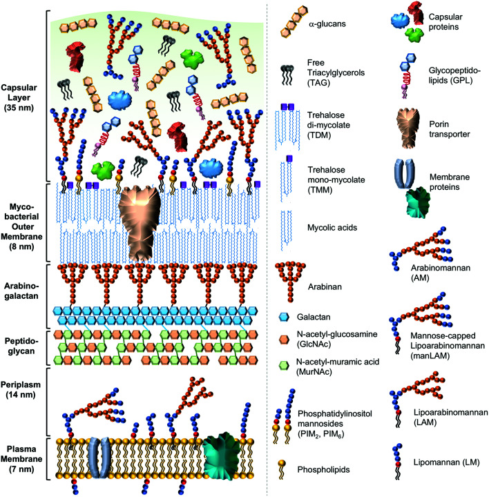
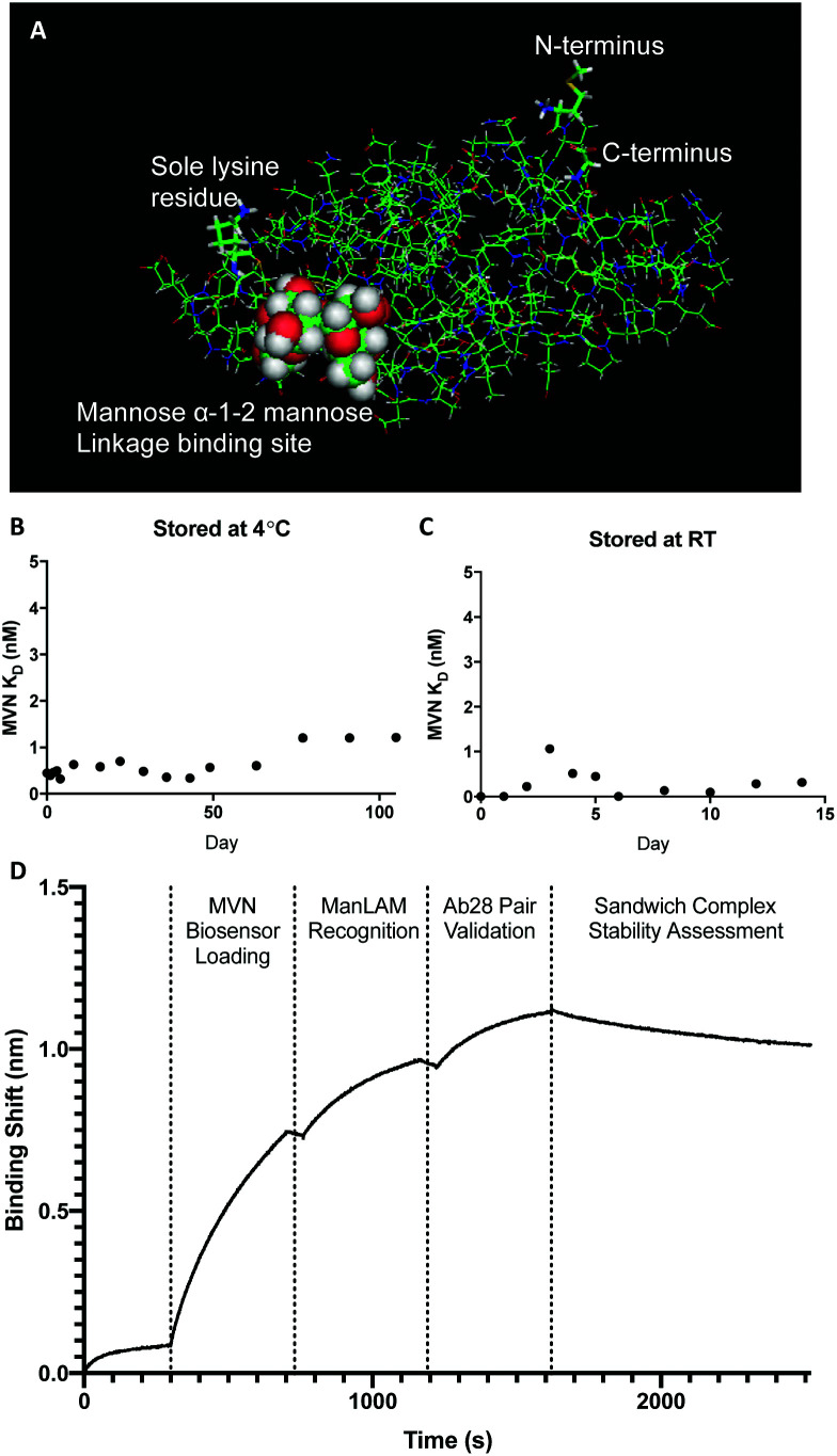
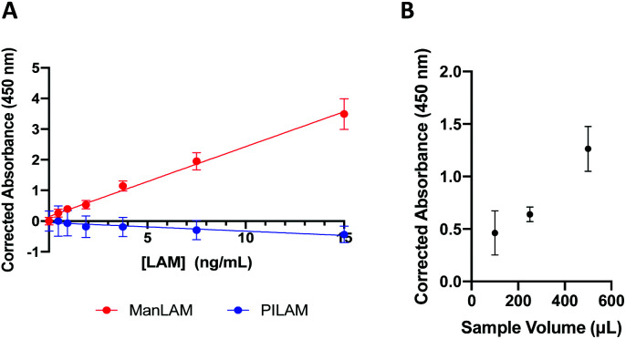
Tuberculosis (TB) is one of the top ten causes of death globally, despite being treatable. The eradication of TB disease requires, amongst others, diagnostic tests with high specificity and sensitivity that will work at the point of care (POC) in low-resource settings. The TB surface glycolipid antigen, mannose-capped lipoarabinomannan (ManLAM) currently serves as the only POC molecular diagnostic biomarker suitable for use in low cost immunoassays. Here, we demonstrate the high affinity and exceptional specificity of microvirin-N (MVN), a 14.3 kDa cyanobacterial lectin, toward H37Rv TB ManLAM and utilize it to develop a novel on-bead ELISA. MVN binds to ManLAM with sub-picomolar binding affinity, but does not bind to other variants of LAM expressed by non-pathogenic mycobacteria - a level of binding specificity and affinity that current commercially available anti-LAM antibodies cannot achieve. An on-bead ELISA was subsequently developed using MVN-functionalized magnetic beads which allows for the specific capture of ManLAM from human urine with a limit of detection (LOD) of 1.14 ng mL-1 and no cross-reactivity when tested with PILAM, a variant of LAM found on non-pathogenic mycobacteria.


Conflict of interest statement

There are no conflicts of interest to declare.

Figures

Fig. 1
Fig. 1. Cross-sectional schematic representation of the mycobacterial cell envelope. The cell envelope of mycobacteria comprises four main layers: (i) an innermost plasma membrane, (ii) a complex of peptidoglycan and arabinogalactan, (iii) a mycobacterial outer membrane (MOM), and (iv) an external capsular layer. The plasma membrane (approximately 7 nm thick) consists of standard membrane lipids such as glycerophospholipids, including phosphatidylinositol mannosides (PIMs), lipomannans (LMs), and lipoarabinomannans (LAMs). Slow-growing, pathogenic mycobacteria, such as Mycobacterium tuberculosis, also express mannose-capped LAM (ManLAM) in which the arabinan branch termini are capped with Manα(1-2)Man linkages. In contrast, fast-growing, non-pathogenic mycobacteria, such as Mycobacterium smegmatis, express PILAM in which the arabinan branch termini are capped with phospho-myo-inositol. A periplasmic space (14 nm) separates the plasma membrane from the innermost layer of the cell wall, the peptidoglycan layer, which serves to maintain cell shape and reinforce the plasma membrane against the osmotic pressure of the cytoplasm. The peptidoglycan layer is composed of linear strands of alternating N-acetyl-glucosamine (GlcNAc) and N-acetyl-muramic acid (MurNAc) sugars that are crosslinked together by peptides into honeycomb-like layers. The peptidoglycan is in turn crosslinked to the middle layer of the cell wall, the arabinogalactan polymer, which is composed of branched arabinose chains and galactose sugars. The mycobacterial outer membrane (MOM) is composed of (a) an inner leaflet of mycolic acids esterified to the underlying arabinogalactan layer, and (b) an outer leaflet composed of both free mycolic acids, and mycolic acids covalently linked to trehalose sugar as either trehalose monomycolate (TMM) or trehalose dimycolate (TDM) variants. The outer leaflet of the mycomembrane also consists of PIMs, LMs, LAMs, and secreted proteins. Porin transporter proteins traverse the entire width of the MOM. Notably, the thickness of the MOM is also similar to that of the plasma membrane (approximately 7–8 nm) despite being composed of very long chain (C60–C90) mycolic acids. The external capsular layer (approximately 35 nm) mainly consists of neutral polysaccharides including a glycogen-like α-glucan and lower amounts of arabinomannan (AM), capsular proteins, and low levels of glycopeptidolipids (GPLs) and free triacylglyercols (TAGs).
Fig. 2
Fig. 2. MVN crystal structure, stability studies, and binding pair assessment. (A) PyMOL rendering of the resolved crystal structure for MVN with the major components labeled. (B) MVN stability study where binding experiments were performed for 100+ days on MVN stored at 4 °C. (C) MVN stability study performed for 2 weeks on MVN stored at RT. (D) FIND Ab28 forms an orthogonal binding pair with MVN.
Fig. 3
Fig. 3. Results of the MVN-based on-bead ELISA. (A) Standard curves of ManLAM and PILAM spiked into pooled human urine. (B) Using larger urine sample volumes in the OB-ELISA allows the resulting signal to be increased proportionally.

Similar articles

Cited by

References

    1. World Health Organization, Global Tuberculosis Report 2018, World Health Organization, S.l., 2018
    1. Pai M. Nicol M. P. Boehme C. C. Microbiol. Spectrum. 2016;4:5. - PubMed
    1. MacLean E. Saravu K. Pai M. Curr. Opin. HIV AIDS. 2019;14(1):46–54. doi: 10.1097/COH.0000000000000512. - DOI - PubMed
    1. Kwan C. K. Ernst J. D. Clin. Microbiol. Rev. 2011;24:351–376. doi: 10.1128/CMR.00042-10. - DOI - PMC - PubMed
    1. Harries A. D. Kumar A. M. V. Diagnostics. 2018;8(4):78. doi: 10.3390/diagnostics8040078. - DOI - PMC - PubMed