. 2021 Feb 12;144(1):128-143.
doi: 10.1093/brain/awaa386.

Vestibular agnosia in traumatic brain injury and its link to imbalance

Affiliations

Vestibular agnosia in traumatic brain injury and its link to imbalance

Elena Calzolari et al. Brain. .

Abstract

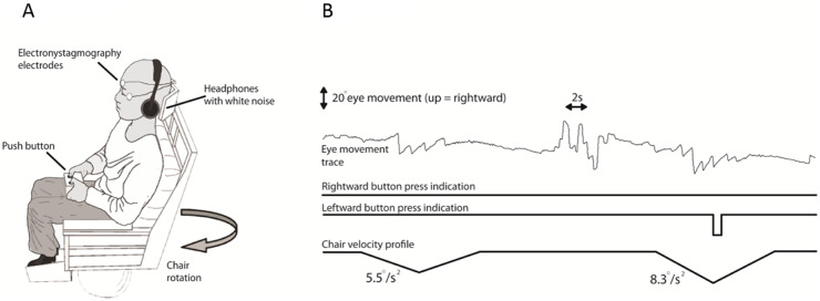
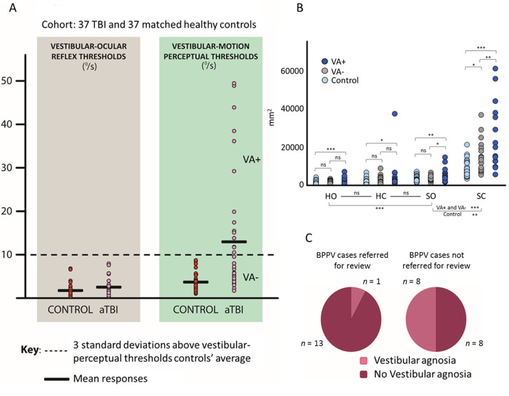
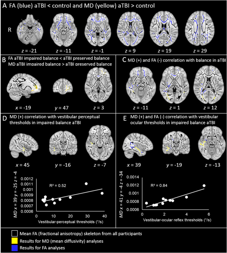
Vestibular dysfunction, causing dizziness and imbalance, is a common yet poorly understood feature in patients with TBI. Damage to the inner ear, nerve, brainstem, cerebellum and cerebral hemispheres may all affect vestibular functioning, hence, a multi-level assessment-from reflex to perception-is required. In a previous report, postural instability was the commonest neurological feature in ambulating acute patients with TBI. During ward assessment, we also frequently observe a loss of vertigo sensation in patients with acute TBI, common inner ear conditions and a related vigorous vestibular-ocular reflex nystagmus, suggesting a 'vestibular agnosia'. Patients with vestibular agnosia were also more unbalanced; however, the link between vestibular agnosia and imbalance was confounded by the presence of inner ear conditions. We investigated the brain mechanisms of imbalance in acute TBI, its link with vestibular agnosia, and potential clinical impact, by prospective laboratory assessment of vestibular function, from reflex to perception, in patients with preserved peripheral vestibular function. Assessment included: vestibular reflex function, vestibular perception by participants' report of their passive yaw rotations in the dark, objective balance via posturography, subjective symptoms via questionnaires, and structural neuroimaging. We prospectively screened 918 acute admissions, assessed 146 and recruited 37. Compared to 37 matched controls, patients showed elevated vestibular-perceptual thresholds (patients 12.92°/s versus 3.87°/s) but normal vestibular-ocular reflex thresholds (patients 2.52°/s versus 1.78°/s). Patients with elevated vestibular-perceptual thresholds [3 standard deviations (SD) above controls' average], were designated as having vestibular agnosia, and displayed worse posturography than non-vestibular-agnosia patients, despite no difference in vestibular symptom scores. Only in patients with impaired postural control (3 SD above controls' mean), whole brain diffusion tensor voxel-wise analysis showed elevated mean diffusivity (and trend lower fractional anisotropy) in the inferior longitudinal fasciculus in the right temporal lobe that correlated with vestibular agnosia severity. Thus, impaired balance and vestibular agnosia are co-localized to the inferior longitudinal fasciculus in the right temporal lobe. Finally, a clinical audit showed a sevenfold reduction in clinician recognition of a common peripheral vestibular condition (benign paroxysmal positional vertigo) in acute patients with clinically apparent vestibular agnosia. That vestibular agnosia patients show worse balance, but without increased dizziness symptoms, explains why clinicians may miss treatable vestibular diagnoses in these patients. In conclusion, vestibular agnosia mediates imbalance in traumatic brain injury both directly via white matter tract damage in the right temporal lobe, and indirectly via reduced clinical recognition of common, treatable vestibular diagnoses.

Keywords: self-motion perception; traumatic brain injury; vertigo; vestibular agnosia; vestibular cognition.


Figures

Figure 1
Figure 1
Vestibular thresholds. Apparatus and methods. (A) Participants sat on a computer-controlled rotating chair (earth-vertical axis). Horizontal eye movements were recorded with electro-nystagmography. Participants indicated their perceived direction of motion by pressing a button to indicate leftward or rightward motion. White noise was delivered through earphones. (B) Raw traces for two subsequent rotations for Patient 08. The top trace shows the electro-nystagmography signal. The middle and bottom traces show the patient’s button press to indicate perceived motion direction, right and left. In this example, the chair rotated from rest to the left, at constant acceleration. The chair continues to accelerate until a correct button response is made or if 5 s has elapsed without a correct button press, or no button press, as here. As no response was made during the test period over 5 s, the chair underwent a controlled deceleration to a stop over 5 s. The second rotation, here also to the left (the rotation directions are randomized), was of increased angular acceleration as determined by the Modified Binary Search (MOBS) algorithm (Tyrrell and Owens, 1988; see Supplementary material for further details). In general, for a given direction (left versus right), a non-perceived rotation is followed by a rotation of higher acceleration, while a perceived rotation is followed by a lower acceleration rotation. Further detail on how the test terminates, and hence thresholds obtained for left and right rotations, can be found in the Supplementary material.
Figure 2
Figure 2
Vestibular threshold testing. (A) Vestibular agnosia in acute TBI. Vestibular-ocular (left) and vestibular-perceptual (right) thresholds to angular acceleration, in healthy controls (dark red) and acute TBI patients (light pink): the acceleration thresholds are displayed in terms of the equivalent instantaneous angular velocity at the time of the threshold detection. Vestibular-ocular thresholds (expressed in degrees per second), correspond to the minimum angular velocity needed to elicit a vestibular-ocular response (first slow-phase of a nystagmus with minimum of two slow and fast phase components). Perceptual thresholds (expressed in degrees per second), correspond to the minimum angular velocity needed to induce the perception of self-motion in the correct direction as assessed by the MOBS procedure. (B) Imbalance in acute TBI assessed via posturography. Sway expressed in square millimetres as the area of the 95% bivariate confidence ellipse of the total displacement of the centre of pressure, in the four posturography conditions (HO = hard surface with eyes open; HC = hard surface with eyes closed; SO = soft surface with eyes open; SC = soft surface with eyes closed), in controls (light blue), acute TBI patients without vestibular agnosia (VA-, grey), and acute TBI patients with vestibular agnosia (VA+, blue). ns = not significant. *P < 0.05; **P < 0.01; ***P < 0.001. (C) Clinically apparent vestibular agnosia masks the presence of BPPV in acute TBI. Left: Patients with BPPV, diagnosed after being referred by the ward clinical staff (n = 14). Right: Patients with BPPV, who were not referred by the ward clinical staff, but diagnosed by our systematic screening on the trauma ward (n = 16). The dark red sectors represent the proportions of patients who reported dizziness during manoeuvres, i.e. they did not have vestibular agnosia. The light pink sectors represent the patients with vestibular agnosia, i.e. they denied dizziness on direct questioning, during manoeuvres that triggered a vestibular nystagmus indicative of BPPV.
Figure 3
Figure 3
Widespread white matter disruption following TBI and correlations with behavioural measures. All contrasts are overlaid upon a standard MNI 152 T1 1 mm brain atlas and the mean FA skeleton (black) with display thresholds set to range from 0.2 to 0.8. The results of FA tract-based spatial statistics contrasts (blue) and the results of MD tract-based spatial statistics contrasts (yellow), are thresholded at P <0.05, corrected for multiple comparisons. (A) Axial slices of the results of the FA contrast acute TBI < control (blue), and of the MD contrast between acute TBI > control groups (yellow). (B) Sagittal, coronal and axial slices of the results of the FA contrast between patients with impaired balance < patients with preserved balance (blue), and of the MD contrast between patients with impaired balance > patients with preserved balance (yellow). (C) Axial slices of the results of the contrast where MD values positively correlate with balance performance (yellow), and where FA values negatively correlate with balance performance in acute TBI patients (i.e. the higher the MD values, the more instability in acute TBI; the lower the FA values, the more instability in acute TBI). (D) Top: Orthogonal view of the areas in the inferior longitudinal fasciculus where patients with impaired balance (but not patients with preserved balance and controls) showed significant positive correlations between MD and vestibular-perceptual thresholds (i.e. the higher the MD value, the more severe the vestibular agnosia, in acute TBI with impaired balance). Bottom: The plot shows the positive correlation between MD and vestibular-perceptual thresholds (°/s) in the significant voxel in the inferior longitudinal fasciculus with the highest correlation with vestibular-perceptual thresholds (x =39, y = −25, z = −4). (E) Top: Orthogonal view of the areas where patients with impaired balance (but not patients with preserved balance and controls) showed significant correlations between vestibular-ocular reflex thresholds and MD (positive correlation, in yellow) and between vestibular-ocular thresholds and FA (negative correlations, in blue). Bottom: For illustrative purposes, the plot shows the positive correlation between MD and vestibular-ocular reflex thresholds (°/s), in the voxel with the highest correlation with vestibular-ocular reflex thresholds (x =41, y = −4, z = −34).

Similar articles

Cited by

References

    1. Agosta F, Galantucci S, Svetel M, Lukić MJ, Copetti M, Davidovic K, et al.Clinical, cognitive, and behavioural correlates of white matter damage in progressive supranuclear palsy. J Neurol 2014; 261: 913–24. - PubMed
    1. Behrens TEJ, Woolrich MW, Jenkinson M, Johansen-Berg H, Nunes RG, Clare S, et al.Characterization and propagation of uncertainty in diffusion-weighted MR imaging. Magn Reson Med 2003; 50: 1077–88. - PubMed
    1. Bonnelle V, Leech R, Kinnunen KM, Ham TE, Beckmann CF, de Boissezon X, et al.Default mode network connectivity predicts sustained attention deficits after traumatic brain injury. J Neurosci 2011; 31: 13442–51. - PMC - PubMed
    1. Brandt T, Strupp M, Benson J.. You are better off running than walking with acute vestibulopathy. Lancet 1999; 354: 746. - PubMed
    1. Chamelian L, Feinstein A.. Outcome after mild to moderate traumatic brain injury: the role of dizziness. Arch Phys Med Rehabil 2004; 85: 1662–6. - PubMed

Publication types

MeSH terms