. 2021 Feb 18;36(3):771-784.
doi: 10.1093/humrep/deaa300.

Mitochondria-targeted therapeutics, MitoQ and BGP-15, reverse aging-associated meiotic spindle defects in mouse and human oocytes

Affiliations

Mitochondria-targeted therapeutics, MitoQ and BGP-15, reverse aging-associated meiotic spindle defects in mouse and human oocytes

Usama Al-Zubaidi et al. Hum Reprod. .

Abstract

Study question: Do mitochondria-targeted therapies reverse ageing- and oxidative stress-induced spindle defects in oocytes from mice and humans?

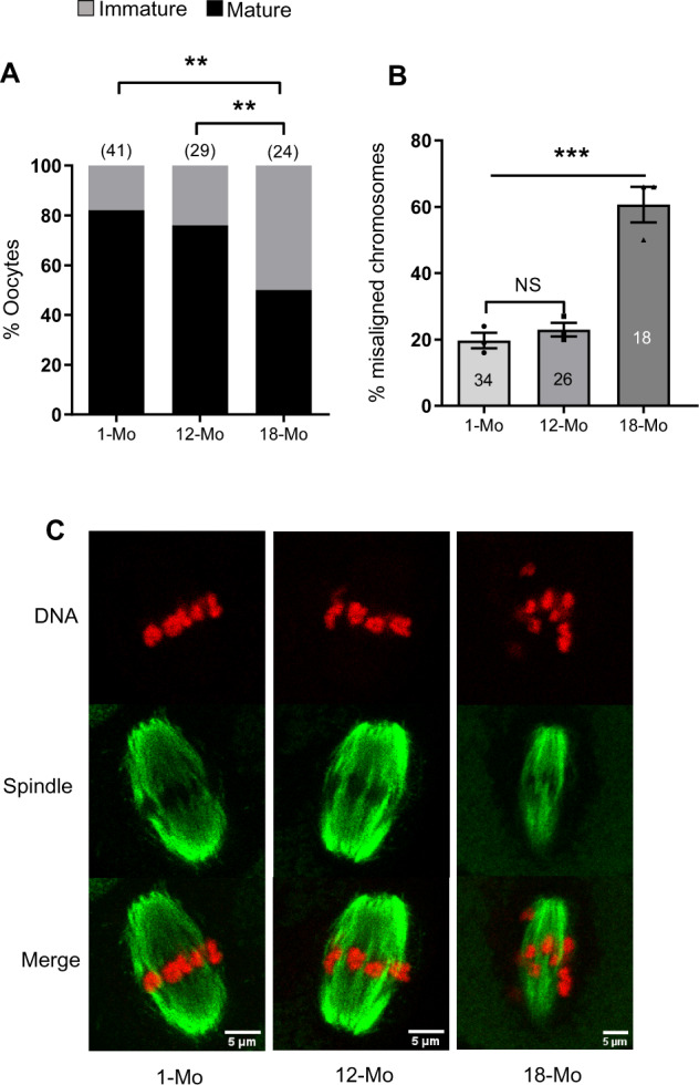
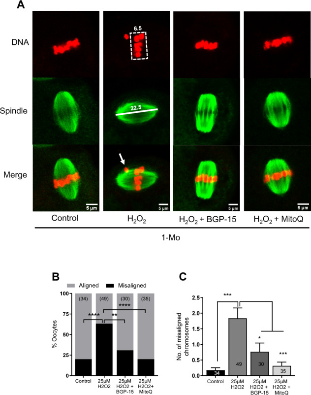
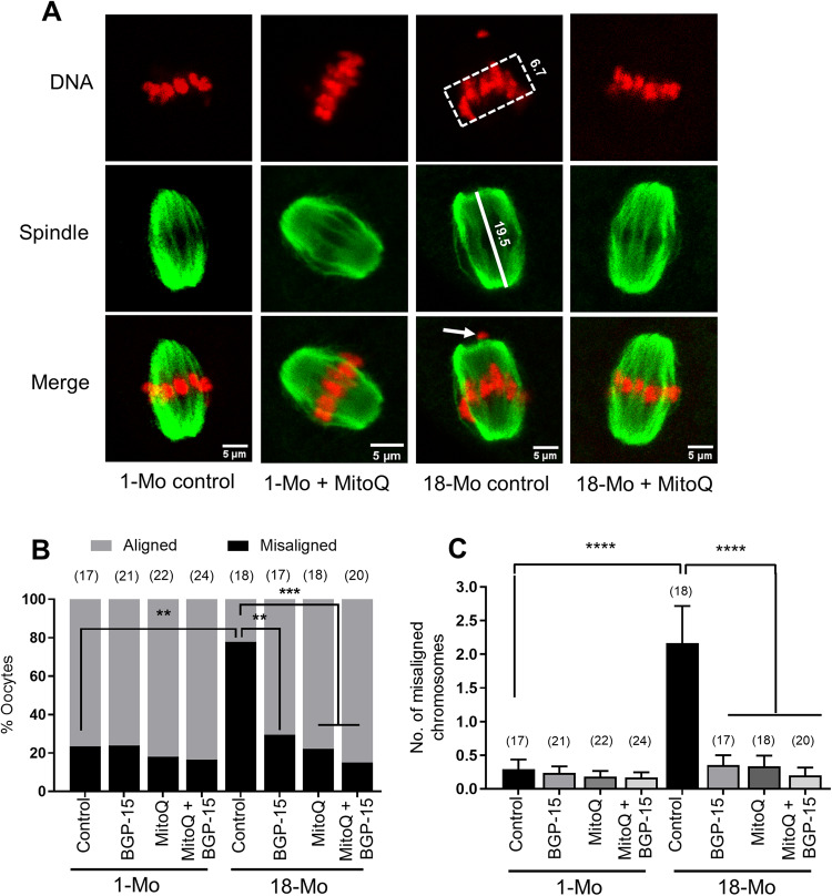
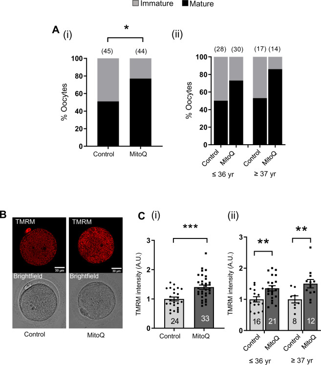
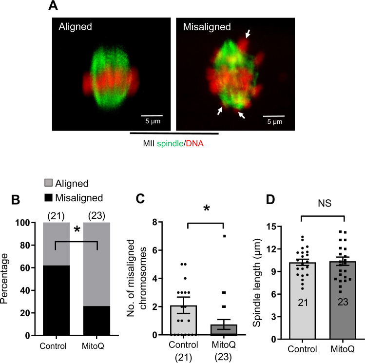
Summary answer: Exposure to MitoQ or BGP-15 during IVM protected against spindle and chromosomal defects in mouse oocytes exposed to oxidative stress or derived from reproductively aged mice whilst MitoQ promoted nuclear maturation and protected against chromosomal misalignments in human oocytes.

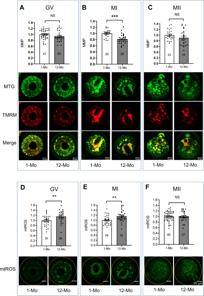
What is known already: Spindle and chromosomal abnormalities in oocytes are more prevalent with maternal aging, increasing the risk of aneuploidy, miscarriage and genetic disorders such as Down's syndrome. The origin of compromised oocyte function may be founded in mitochondrial dysfunction and increased reactive oxygen species (ROS).

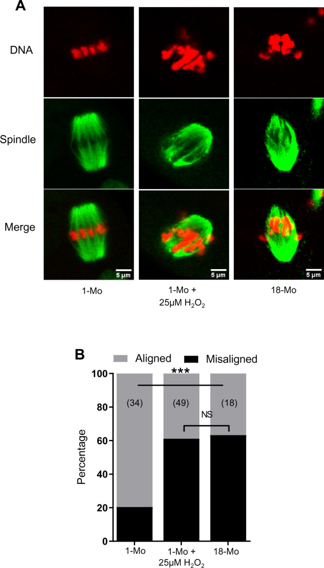
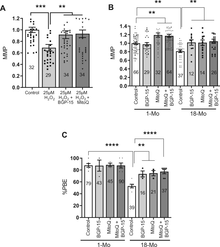
Study design, size, duration: Oocytes from young and old mice were treated with MitoQ and/or BGP-15 during IVM. To directly induce mitochondrial dysfunction, oocytes were treated with H2O2, and then treated the MitoQ and/or BGP-15. Immature human oocytes were cultured with or without MitoQ. Each experiment was repeated at least three times, and data were analyzed by unpaired-sample t-test or chi-square test.

Participants/materials, setting, methods: Immature germinal vesicle (GV) stage oocytes from 1-, 12- and 18-month-old mice were obtained from preovulatory ovarian follicles. Oocytes were treated with MitoQ and/or BGP-15 during IVM. GV-stage human oocytes were cultured with or without MitoQ. Mitochondrial membrane potential and mitochondrial ROS were measured by live-cell imaging. Meiotic spindle and chromosome alignments were visualized by immunofluorescent labeling of fixed oocytes and the 3-dimensional images were analyzed by Imaris.

Main results and the role of chance: MitoQ or BGP-15 during IVM protects against spindle and chromosomal defects in oocytes exposed to oxidative stress and in oocytes from aged mice (P < 0.001). In human oocytes, the presence of MitoQ during IVM promoted nuclear maturation and had a similar positive effect in protecting against chromosomal misalignments (P < 0.001).

Limitations, reasons for caution: Our study identifies two excellent candidates that may help to improve fertility in older women. However, these potential therapies must be tested for efficacy in clinical IVM systems, and undergo thorough examination of resultant offspring in preclinical models before utilization.

Wider implications of the findings: Our results using in-vitro systems for oocyte maturation in both mouse and human provide proof of principle that mitochondrially targeted molecules such as MitoQ and BGP-15 may represent a novel therapeutic approach against maternal aging-related spindle and chromosomal abnormalities.

Study funding/competing interest(s): The project was financially supported by the National Health and Medical Research Council and Australian Research Council, Australia. U.A.-Z. was supported by the Iraqi Higher Education and Scientific Research Ministry PhD scholarship and O.C. was supported by TUBITAK-1059B191601275. M.P.M. consults for MitoQ Inc. and holds patents in mitochondria-targeted therapies. R.L.R. is an inventor on patents relating to the use of BGP-15 to improve gamete quality.

Trial registration number: N/A.

Keywords: aging; chromosome; mitochondria-targeted therapeutics; oocyte; spindle.


Figures

Figure 1.
Figure 1.
Maturation and chromosomal misalignment rates of oocytes isolated from mice at different ages. (A) Maturation rates after in-vitro culture. (B) Percentage of oocytes with one or more misaligned chromosomes. (C) Representative images of MII oocytes with labeled spindles (green) and DNA (red). Mo, month. Results are presented from at least three replicate experiments in all cases. Error bars show SE of the mean. Number of oocytes analyzed in each group are shown. **P ≤ 0.01; ***P ≤ 0.001 and NS, not significant.
Figure 2
Figure 2
Comparison of mitochondrial membrane potential and mitochondrial reactive oxygen species (ROS) during oocyte maturation. Germinal vesicle, and metaphase I and II (GV, MI and MII) stage oocytes were labeled with Tetramethylrhodamine, Methyl Ester, Perchlorate (TMRM) and mitotracker green (MTG) and mitochondrial membrane potential (MMP) was calculated as the ratio of TMRM and MTG. (A) MMP comparison in GV-stage oocytes. (B) MMP comparison in MI-stage oocytes. (C) MMP comparison in MII-stage oocytes. Corresponding representative images of oocytes labeled with MTG (green), TMRM (red) and merged (yellow) are shown under each figure. GV, MI and MII oocytes from young and 12-month-old mice were labeled with MitoSOX red. (D) Comparison of ROS in GV-stage oocytes. (E) Comparison of ROS in MI-stage oocytes. (F) Comparison of ROS in MII-stage oocytes. Representative images of MitoSOX-labeled (green) oocytes are shown below each panel. Error bars show SE of the mean. Number of oocytes analyzed in each group are shown. **P ≤ 0.01; ***P ≤ 0.001 and NS, not significant.
Figure 3.
Figure 3.
Generation of reactive oxygen species (ROS)-induced model of spindle and chromosomal defects. Germinal vesicle (GV) stage oocytes were collected from 1 month (1-Mo) and 18-Mo-old mice and were matured in vitro in M2 medium. (A) Metaphase II (MII) oocytes in 1-Mo group either remained in M2 medium or were treated with 25 µM H2O2 for 1 h. The 18-Mo group MII oocytes remained untreated. Mature oocytes were fixed and labeled for spindles (green) and DNA (red). (B) Percentage of oocytes containing aligned or misaligned chromosomes in different groups. Number of oocytes analyzed in each group are shown. ***P ≤ 0.001 and NS, not significant.
Figure 4.
Figure 4.
Enhanced mitochondrial membrane potential (MMP) and oocyte maturation rates by mitochondria-targeted agents. (A) H2O2 significantly decreased MMP but BGP-15 or MitoQ can prevent such decrease in MMP in metaphase II (MII) oocytes from 1 month (1-Mo) old mice. (B) Comparison MMP in MII oocytes from young (1-Mo) and old (18-Mo) mice after IVM in the absence or presence of BGP-15, MitoQ and both combined. Number of oocytes analyzed in each group are shown. (C) Comparison of maturation rates of oocytes from young (1-Mo) and old (18-Mo) mice after in-vitro culture in the absence or presence of BGP-15, MitoQ and both combined. Error bars show SE of the mean. Number of oocytes analyzed in each group are shown. **P ≤ 0.01, ****P ≤ 0.0001.
Figure 5.
Figure 5.
Rescue of chromosomal misalignments induced by oxidative stress in young mouse oocytes. (A) Germinal vesicle (GV) stage oocytes collected from 1 month (1-Mo) old mice were matured in M2 medium only (control) or in the presence of 10 µM BGP-15 or 50 nM MitoQ. Metaphase II (MII) oocytes were treated with 25 µM H2O2 for 1 h and were fixed and labeled for spindles (green) and DNA (red). (B) Percentages of oocytes with aligned or misaligned chromosomes. (C) Quantification of average number of misaligned chromosomes per oocyte in different treatment groups. Error bars show SE of the mean. Number of oocytes analyzed in each group are shown. *P < 0.05; **P < 0.01; ***P < 0.001; ****P < 0.0001.
Figure 6.
Figure 6.
Rescue of chromosomal misalignments in oocytes isolated from aged mice. (A) Germinal vesicle (GV) stage oocytes from 1-month-old and 18-month-old mice were matured in M16 medium only (control) or with 10 µM BGP-15, 50 nM MitoQ or BGP-15 + MitoQ at 37°C with 5% CO2. Mature oocytes were fixed and labeled for spindles and DNA. Representative MII spindle and DNA in control oocyte from 1-Mo-old mice, 1-Mo MitoQ treated, 18-Mo control and 18-Mo MitoQ treated are shown. (B) Percentages of oocytes with aligned or misaligned chromosomes. (C) Quantification of average number of misaligned chromosomes per oocyte. Error bars show SE of the mean. Number of oocytes analyzed in each group are shown. *P < 0.05; **P < 0.01; ***P < 0.001; ****P < 0.0001.
Figure 7.
Figure 7.
IVM of human oocytes in the presence of MitoQ. (A) Comparison of maturation rates between control and 50 nM MitoQ-treated groups (i). Data are expressed as percentages and chi-square test was used to compare between groups, *P < 0.05. (ii) Comparison of maturation rates between control and MitoQ-treated oocytes from (i) in different age groups. (B) Representative Tetramethylrhodamine, Methyl Ester, Perchlorate (TMRM) fluorescence and bright field images. (C) Quantification of TMRM signal intensities between control and MitoQ-treated metaphase II (MII) oocytes (i). (ii) Comparison of TMRM signal intensities between control and MitoQ-treated oocytes from (i) in different age groups. Data are expressed as mean ± SEM. Chi-square test (A) or Student’s t-test (C) was used to compare between groups.; *P < 0.05, **P < 0.01 and ***P < 0.001.
Figure 8.
Figure 8.
Spindle and chromosome alignments in in-vitro matured human oocytes in the presence of MitoQ. (A) Examples of aligned and misaligned chromosomes (arrows). Metaphase II spindles and chromosomes were visualized by staining tubulin and DNA to analyze chromosomal misalignment. (B) Percentage of mature oocytes with aligned and misaligned chromosomes in control and MitoQ groups. (C) Number of misaligned chromosomes per oocyte in control and MitoQ groups. (D) Comparison of spindle length in control and MitoQ groups. Error bars show SE of the mean. Number of oocytes analyzed in each group are shown. Chi-square test (B) or Student’s t-test (C and D) was used to compare between groups. *P < 0.05, NS, not significant.

Similar articles

Cited by

References

    1. Al-Zubaidi U, Liu J, Cinar O, Robker RL, Adhikari D, Carroll J.. The spatio-temporal dynamics of mitochondrial membrane potential during oocyte maturation. Mol Hum Reprod 2019;25:695–705. - PMC - PubMed
    1. Angell RR. Aneuploidy in older women. Higher rates of aneuploidy in oocytes from older women. Hum Reprod 1994;9:1199–1200. - PubMed
    1. Babayev E, Wang T, Szigeti-Buck K, Lowther K, Taylor HS, Horvath T, Seli E.. Reproductive aging is associated with changes in oocyte mitochondrial dynamics, function, and mtDNA quantity. Maturitas 2016;93:121–130. - PMC - PubMed
    1. Baird DT, Collins J, Egozcue J, Evers LH, Gianaroli L, Leridon H, Sunde A, Templeton A, Van Steirteghem A, Cohen J. et al.; ESHRE Capri Workshop Group. Fertility and ageing. Hum Reprod Update 2005;11:261–276. - PubMed
    1. Ben-Meir A, Burstein E, Borrego-Alvarez A, Chong J, Wong E, Yavorska T, Naranian T, Chi M, Wang Y, Bentov Y. et al. Coenzyme Q10 restores oocyte mitochondrial function and fertility during reproductive aging. Aging Cell 2015;14:887–895. - PMC - PubMed

Publication types