. 2021 Mar 25;106(4):e1763-e1774.
doi: 10.1210/clinem/dgaa958.

HLA Class I Upregulation and Antiviral Immune Responses in Graves Disease

Affiliations

HLA Class I Upregulation and Antiviral Immune Responses in Graves Disease

Therese Weider et al. J Clin Endocrinol Metab. .

Abstract

Context: The origin of Graves disease (GD) remains elusive. However, evidence of an association between GD and viral infections is emerging. Human leukocyte antigen (HLA) class I presents viral antigens to circulating immune cells and plays a crucial role in the defense against viral infections.

Objective: This work aimed to investigate HLA class I expression, enterovirus presence, and the viral immune response proteins signal transducer and activation of transcription 1 (STAT1) and protein kinase R (PKR) in thyroid tissue from GD patients.

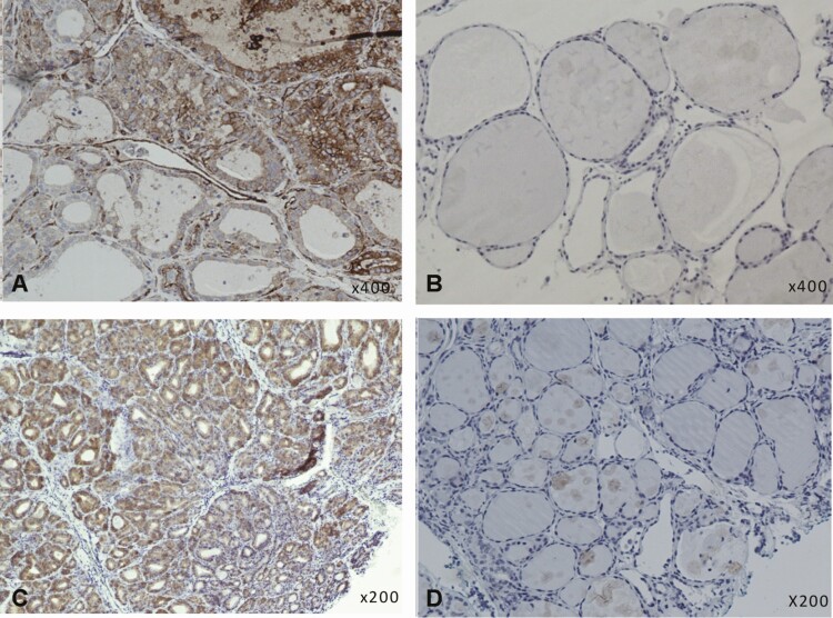
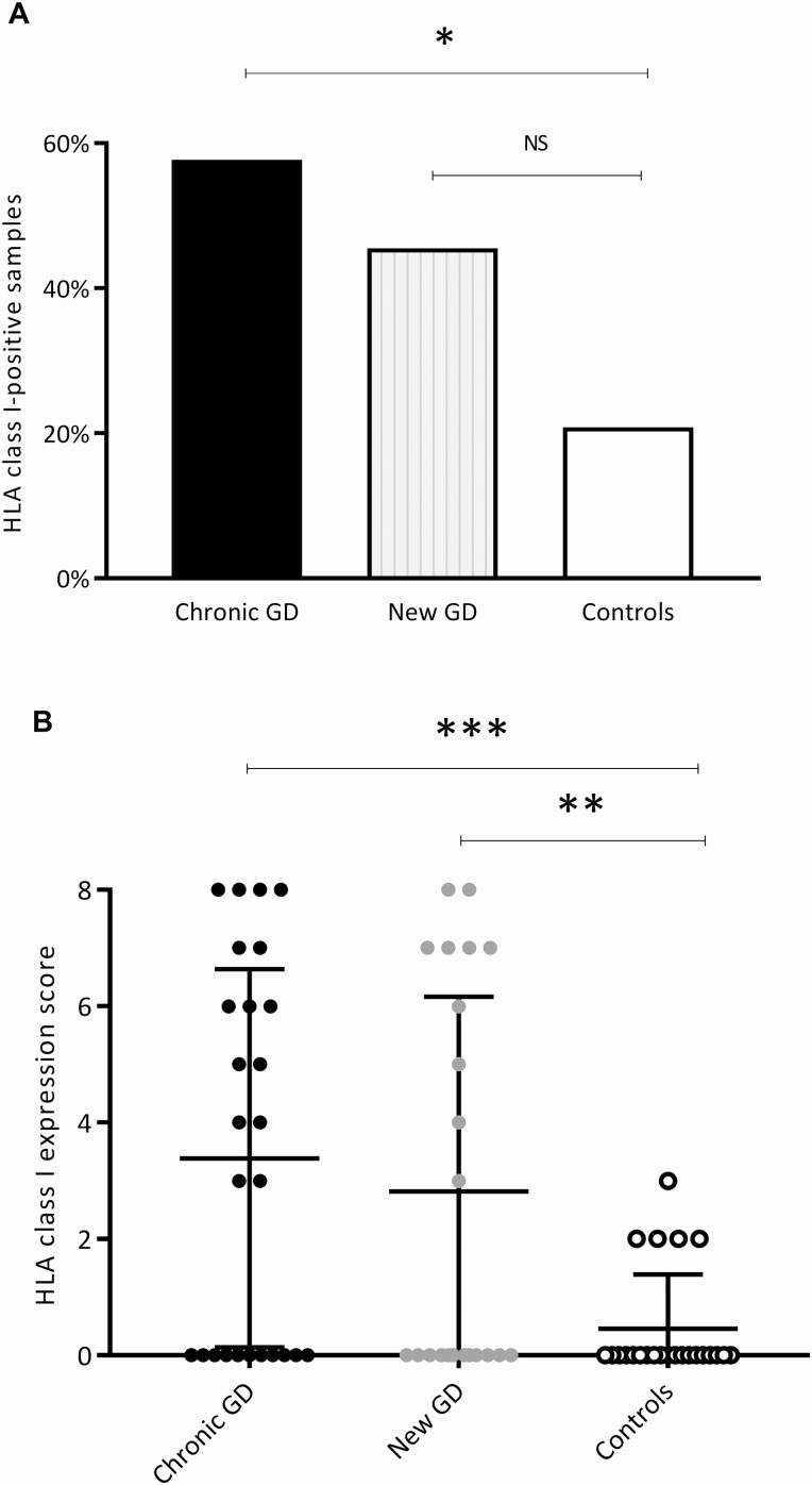
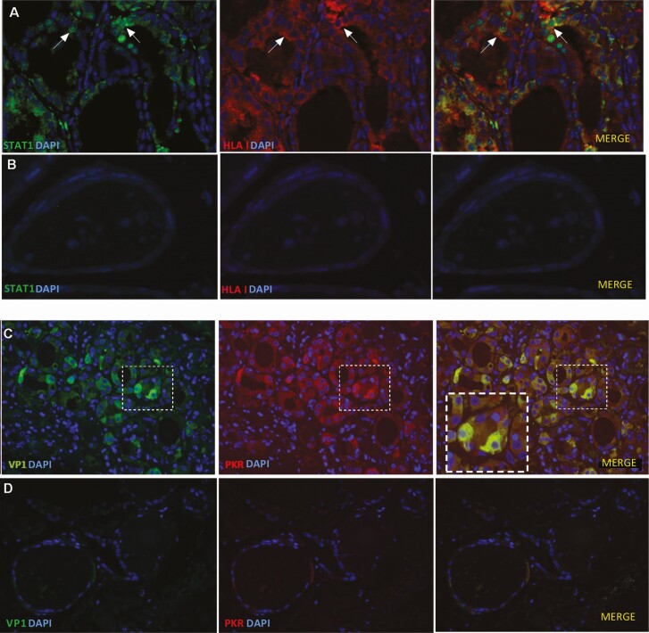
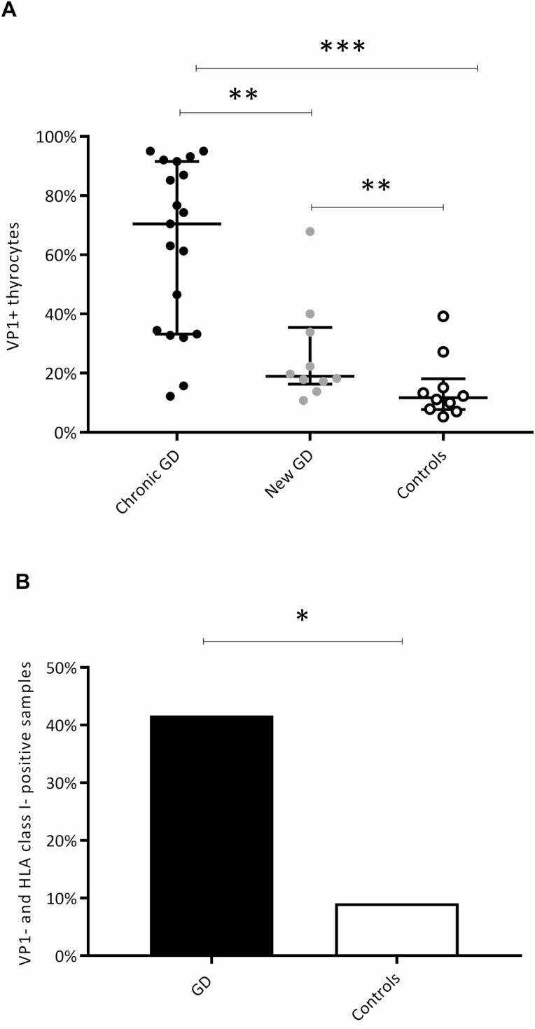
Methods: We collected thyroid tissue from core needle biopsies or surgical specimens from 48 GD patients and 24 controls. Standard immunohistochemistry was used to detect HLA class I and enteroviral capsid protein 1 (VP1) on formalin-fixed and paraffin-embedded tissue. STAT1 and PKR were examined by combined immunofluorescence staining. HLA class I expression score was the main outcome measure.

Results: The HLA class I expression score, which takes both proportion and intensity of immunostaining into account, was significantly higher in GD patients (3.1 ± 3.3) than in controls (0.5 ± 0.9) (P < .001). Significantly more VP1 positive thyroid cells were found GD samples (50.1 ± 30.5%) than in controls (14.9 ± 10.5%) (P < .001). STAT1 and HLA class I were found within the same thyroid cells and PKR and VP1 were also colocalized within thyroid cells.

Conclusion: HLA class I is upregulated in GD and enterovirus protein is prevalent in thyroid tissue. The colocalization of HLA class I with STAT1 and VP1 with PKR indicates an antiviral tissue response. These findings support the concept of a link between viral infections and GD.

Keywords: Graves disease; HLA class I; STAT1; autoimmune thyroid disease; enterovirus; viral infections.


Figures

Figure 1.
Figure 1.
Immunohistochemical staining for HLA class I and VP1 in thyroid tissue samples from GD patients and controls. HLA class I and VP1 was found in more GD samples than controls. A, Positive HLA class I staining (brown color) in thyroid follicular cells in a new GD sample. B, Negative HLA class I staining in a control sample. C, Globally positive VP1 staining (brown color) in thyroid follicular cells in a chronic GD sample. D, Negative VP1 staining in a control sample. All samples stained with a standard horseradish peroxidase immunohistochemistry protocol. GD, Graves disease; HLA, human leukocyte antigen; VP1, enteroviral capsid protein 1.
Figure 2.
Figure 2.
HLA class I immunodetection and HLA class I expression score. Significantly more HLA class I–positive samples were found in the GD group than in the control group. A, Proportion of samples with HLA class I positivity in the 2 GD subgroups and in the controls. B, HLA class I expression score (Allred score), taking both immunostaining intensity and proportion of stained tissue into account (maximum score 8, and lowest score 0). The HLA class I expression score was significantly higher in the chronic GD group and in the newly diagnosed GD group than in controls. Bars represent mean and SD. *P less than or equal to .05; **P less than or equal to .01; *** P less than or equal to .001. GD, Graves disease; HLA, human leukocyte antigen; NS, not significant.
Figure 3.
Figure 3.
Combined immunofluorescence of HLA class I/STAT1 and PKR/VP1 in GD samples and controls. Nuclear and cytosolic STAT1 was found in thyroid cells and was colocalized with HLA class I. PKR and VP1 were also colocalized within thyroid cells. A, An example of STAT1 (green) and HLA class I (red) immunofluorescence staining in GD thyroid tissue. Arrows indicate thyroid cells with nuclear STAT1 and intracellular HLA class I. B, Control sample with negative STAT1 and HLA class I staining. C, An example of VP1 (green) and PKR (red) immunofluorescence staining in GD thyroid tissue. The insets represent higher magnification (of the area outlined by the white boxes) and shows colocalized VP1 and PKR within thyrocytes. D, Control sample with negative VP1 and PKR staining. Merged images with blue nuclear DAPI staining. All scale bars at 25 μm. DAPI, 4’,6-diamidino-2-phenylindole; GD, Graves disease; HLA, human leukocyte antigen; PKR, protein kinase R; STAT1, signal transducer and activator of transcription 1; VP1, enteroviral capsid protein 1.
Figure 4.
Figure 4.
VP1 assessment in GD samples and controls. We confirmed the presence of VP1 in GD samples. A, Number of VP1+ thyrocytes in the GD subgroups and controls. There were significantly more VP1+ thyrocytes both in the chronic GD group and the new GD group compared to controls. The chronic GD group had significantly more VP1+ thyrocytes than the new GD group. Bars represent median and interquartile range. B, Proportion of double-positive samples (VP1+/HLA I+) in the GD group and the controls. We found significantly more double-positive samples in the GD group than in the control group. *P less than or equal to .05; **P less than or equal to .01; ***P less than or equal to .001. GD, Graves disease; HLA, human leukocyte antigen; VP1, enteroviral capsid protein 1.

References

    1. Lee HJ, Li CW, Hammerstad SS, Stefan M, Tomer Y. Immunogenetics of autoimmune thyroid diseases: a comprehensive review. J Autoimmun. 2015;64:82-90. - PMC - PubMed
    1. Chen QY, Huang W, She JX, Baxter F, Volpe R, Maclaren NK. HLA-DRB1*08, DRB1*03/DRB3*0101, and DRB3*0202 are susceptibility genes for Graves’ disease in North American Caucasians, whereas DRB1*07 is protective. J Clin Endocrinol Metab. 1999;84(9):3182-3186. - PubMed
    1. Misaki T, Iida Y, Kasagi K, Konishi J. Seasonal variation in relapse rate of Graves’ disease after thionamide drug treatment. Endocr J. 2003;50(6):669-672. - PubMed
    1. Hammerstad SS, Stefan M, Blackard J, et al. . Hepatitis C virus E2 protein induces upregulation of IL-8 pathways and production of heat shock proteins in human thyroid cells. J Clin Endocrinol Metab. 2017;102(2):689-697. - PMC - PubMed
    1. Desailloud R, Goffard A, Page C, et al. . Detection of enterovirus RNA in postoperative thyroid tissue specimens. Clin Endocrinol (Oxf). 2009;70(2):331-334. - PubMed

Publication types

Substances