

A Walnut Diet in Combination with Enriched Environment Improves Cognitive Function and Affects Lipid Metabolites in Brain and Liver of Aged NMRI Mice
- PMID: 33367957
- PMCID: PMC7929966
- DOI: 10.1007/s12017-020-08639-7
A Walnut Diet in Combination with Enriched Environment Improves Cognitive Function and Affects Lipid Metabolites in Brain and Liver of Aged NMRI Mice
Abstract
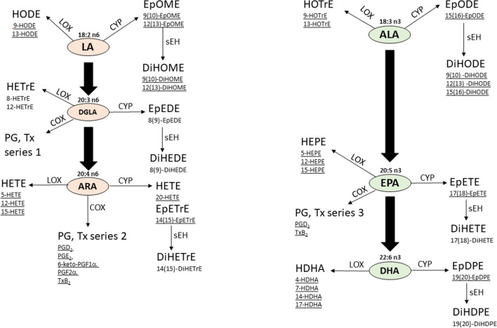
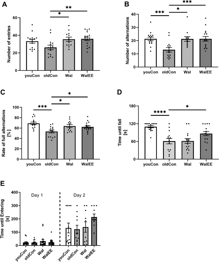
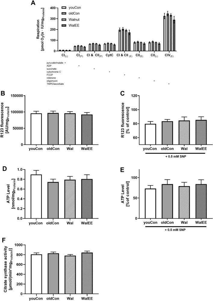
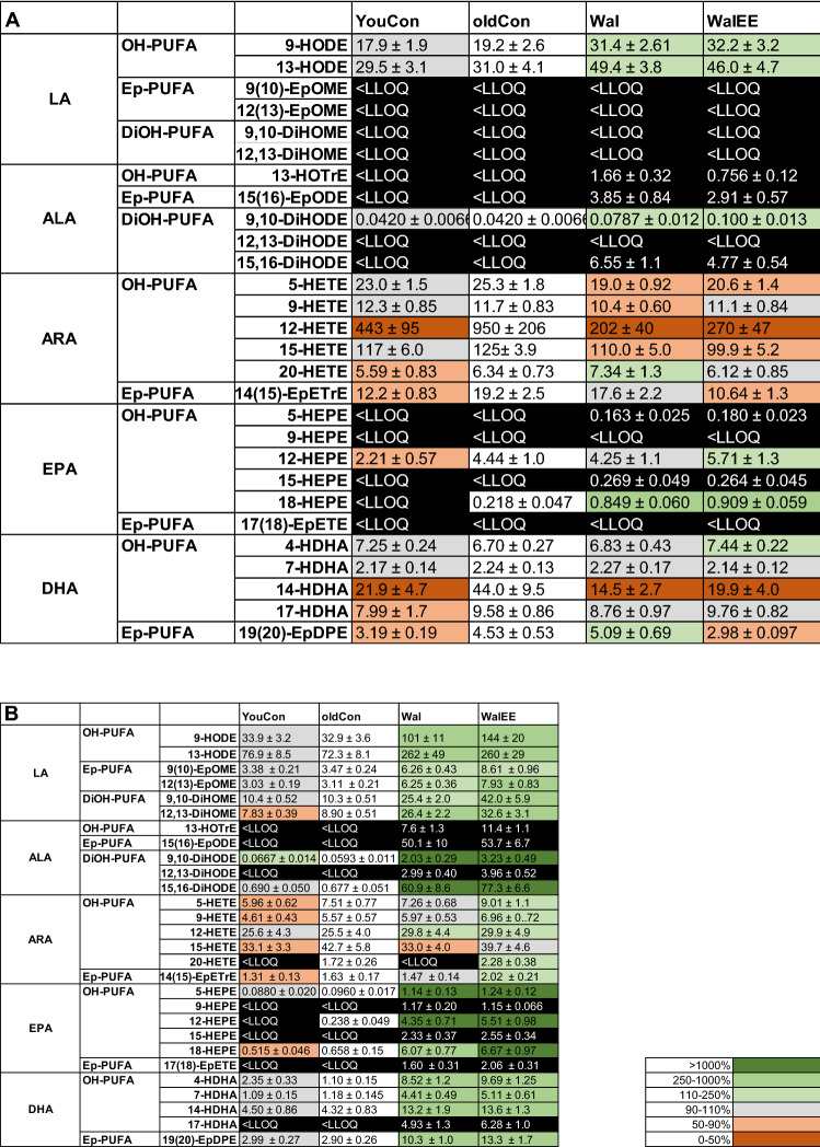
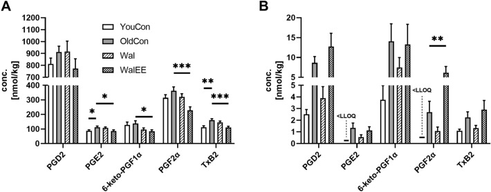
This in vivo study aimed to test if a diet enriched with 6% walnuts alone or in combination with physical activity supports healthy ageing by changing the oxylipin profile in brain and liver, improving motor function, cognition, and cerebral mitochondrial function. Female NMRI mice were fed a 6% walnut diet starting at an age of 12 months for 24 weeks. One group was additionally maintained in an enriched environment, one group without intervention served as control. After three months, one additional control group of young mice (3 weeks old) was introduced. Motor and cognitive functions were measured using Open Field, Y-Maze, Rotarod and Passive Avoidance tests. Lipid metabolite profiles were determined using RP-LC-ESI(-)-MS/MS in brain and liver tissues of mice. Cerebral mitochondrial function was characterized by the determination of ATP levels, mitochondrial membrane potential and mitochondrial respiration. Expression of genes involved with mito- and neurogenesis, inflammation, and synaptic plasticity were determined using qRT-PCR. A 6% walnut-enriched diet alone improved spatial memory in a Y-Maze alternation test (p < 0.05) in mice. Additional physical enrichment enhanced the significance, although the overall benefit was virtually identical. Instead, physical enrichment improved motor performance in a Rotarod experiment (p* < 0.05) which was unaffected by walnuts alone. Bioactive oxylipins like hydroxy-polyunsaturated fatty acids (OH-PUFA) derived from linoleic acid (LA) were significantly increased in brain (p** < 0.01) and liver (p*** < 0.0001) compared to control mice, while OH-PUFA of α-linolenic acid (ALA) could only be detected in the brains of mice fed with walnuts. In the brain, walnuts combined with physical activity reduced arachidonic acid (ARA)-based oxylipin levels (p < 0.05). Effects of walnut lipids were not linked to mitochondrial function, as ATP production, mitochondrial membrane potential and mitochondrial respiration were unaffected. Furthermore, common markers for synaptic plasticity and neuronal growth, key genes in the regulation of cytoprotective response to oxidative stress and neuronal growth were unaffected. Taken together, walnuts change the oxylipin profile in liver and brain, which could have beneficial effects for healthy ageing, an effect that can be further enhanced with an active lifestyle. Further studies may focus on specific nutrient lipids that potentially provide preventive effects in the brain.
Keywords: Ageing; Behaviour; Brain; Cognition; Liver; Mitochondrial function; NMRI; Neurodegeneration; Oxylipins.
Conflict of interest statement
The authors declare no conflict of interest.
Figures
Similar articles
-
A Walnut-Enriched Diet for 2 Years Changes the Serum Oxylipin Profile in Healthy Older Persons.J Nutr. 2024 Feb;154(2):395-402. doi: 10.1016/j.tjnut.2023.12.007. Epub 2023 Dec 9. J Nutr. 2024. PMID: 38081585 Clinical Trial.
-
Beneficial Effects of Walnuts on Cognition and Brain Health.Nutrients. 2020 Feb 20;12(2):550. doi: 10.3390/nu12020550. Nutrients. 2020. PMID: 32093220 Free PMC article. Review.
-
The Brain Oxylipin Profile Is Resistant to Modulation by Dietary n-6 and n-3 Polyunsaturated Fatty Acids in Male and Female Rats.Lipids. 2019 Jan;54(1):67-80. doi: 10.1002/lipd.12122. Epub 2019 Jan 29. Lipids. 2019. PMID: 30697757
-
Dietary supplementation of walnuts improves memory deficits and learning skills in transgenic mouse model of Alzheimer's disease.J Alzheimers Dis. 2014;42(4):1397-405. doi: 10.3233/JAD-140675. J Alzheimers Dis. 2014. PMID: 25024344
-
Long-chain polyunsaturated fatty acids (LCPUFA) from genesis to senescence: the influence of LCPUFA on neural development, aging, and neurodegeneration.Prog Lipid Res. 2014 Jan;53:1-17. doi: 10.1016/j.plipres.2013.10.002. Epub 2013 Oct 24. Prog Lipid Res. 2014. PMID: 24334113 Review.
Cited by
-
Walnut Oil Reduces Aβ Levels and Increases Neurite Length in a Cellular Model of Early Alzheimer Disease.Nutrients. 2022 Apr 19;14(9):1694. doi: 10.3390/nu14091694. Nutrients. 2022. PMID: 35565661 Free PMC article.
-
Neuroprotective effects of walnut (Juglans regia L.) in nervous system disorders: A comprehensive review.Iran J Basic Med Sci. 2024;27(12):1492-1505. doi: 10.22038/ijbms.2024.79854.17302. Iran J Basic Med Sci. 2024. PMID: 39539440 Free PMC article. Review.
-
The Role of Alpha-Linolenic Acid and Other Polyunsaturated Fatty Acids in Mental Health: A Narrative Review.Int J Mol Sci. 2024 Nov 20;25(22):12479. doi: 10.3390/ijms252212479. Int J Mol Sci. 2024. PMID: 39596544 Free PMC article. Review.
-
Impact of aging on mitochondrial respiration in various organs.Physiol Res. 2022 Dec 31;71(S2):S227-S236. doi: 10.33549/physiolres.934995. Physiol Res. 2022. PMID: 36647911 Free PMC article.
-
Caenorhabditis elegans as a Model for the Effects of Phytochemicals on Mitochondria and Aging.Biomolecules. 2022 Oct 24;12(11):1550. doi: 10.3390/biom12111550. Biomolecules. 2022. PMID: 36358900 Free PMC article. Review.
References
-
- Afshordel S, Hagl S, Werner D, Röhner N, Kögel D, Bazan NG, et al. Omega-3 polyunsaturated fatty acids improve mitochondrial dysfunction in brain aging–impact of Bcl-2 and NPD-1 like metabolites. Prostaglandins, leukotrienes, and essential fatty acids. 2015;92:23–31. doi: 10.1016/j.plefa.2014.05.008. - DOI - PubMed
Publication types
MeSH terms
Substances
LinkOut - more resources
Full Text Sources
Medical
