Review
. 2021 Mar;8(1):63-80.
doi: 10.1007/s40744-020-00264-x. Epub 2020 Dec 24.

Renal Disease in Primary Sjögren's Syndrome

Affiliations
Review

Renal Disease in Primary Sjögren's Syndrome

Oshorenua Aiyegbusi et al. Rheumatol Ther. 2021 Mar.

Abstract

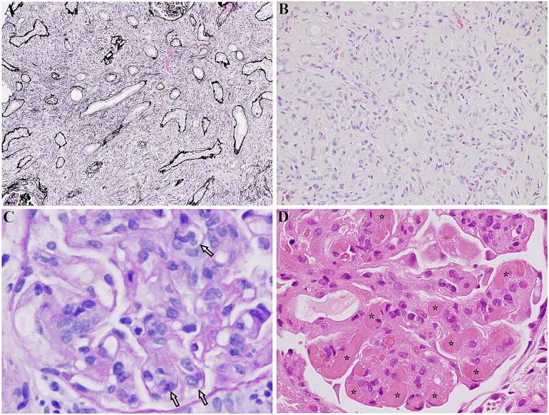
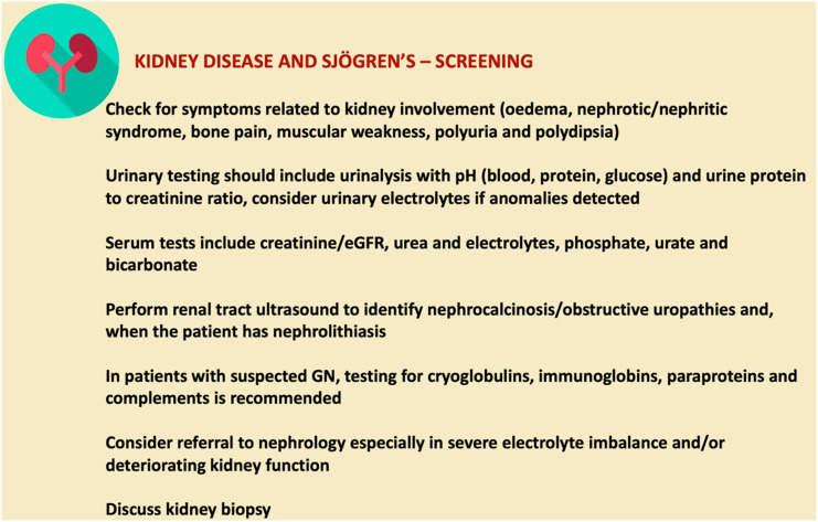
Primary Sjögren's syndrome (pSS) is a chronic autoimmune disorder characterised by lymphocytic infiltration of the exocrine glands, predominantly the salivary and lacrimal glands, leading to sicca symptoms. Patients may have extraglandular disease involving multiple organs, including the kidneys. 5% of patients with pSS can have renal involvement. Kidney disease in pSS presents a diagnostic challenge, as clinical symptoms are often insidious and can precede sicca symptoms. pSS affects the kidney through lymphocytic infiltration of renal tubules or immune complex deposition, leading to an array of clinical features. Tubulointerstitial nephritis is the most common histological pattern of kidney disease. Other tubular injuries include renal tubular acidosis with hypokalaemia, Fanconi's syndrome and diabetes insipidus. Glomerular disease is less common and typically involves an immune complex-mediated process. Optimal treatment for kidney diseases in pSS is not established, and treatment is guided by the pattern of disease. For tubulointerstitial nephritis, management involves electrolyte imbalance correction and the use of immunosuppression, including steroids. Treatment of glomerular disease is targeted to the histological pattern, and often requires a combination of immunosuppressive agents. The risk of end-stage kidney disease is low. Nevertheless, patients with pSS and kidney disease have significantly reduced quality of life.

Keywords: Glomerulonephritis; Kidney disease; Sjögren’s syndrome; Tubular dysfunction; Tubulointerstitial nephritis.


Figures

Fig. 1
Fig. 1
Distal tubule. The α-intercalated cell is responsible for H+ secretion by H+/ATPase and H+/K+/ATPase. Ammonia (NH3) buffers H+ to form ammonium in the lumen. Intracellularly, HCO3 leaves cell via Cl/HCO3 exchange facilitated by AE1 (anion exchanger). Carbonic anhydrase II (CA II) is needed to secrete H+
Fig. 2
Fig. 2
Proximal tubule. Intracellular carbonic acid (H2CO3) dissociates into H+ and HCO3 under the action of carbonic anhydrase II (CAII). H+ secretion is facilitated by Na+/H+ exchanger, and Na+/HCO3 cotransporter is responsible for HCO3 transport. In the lumen, H+ reacts with HCO3 to form H2CO3, which dissociates into H2O and CO2 through the action of carbonic anhydrase V (CA V). Glucose, amino acids, phosphate and other substances are also reabsorbed through active and passive processes in the proximal tubule (mechanism not shown in schematic diagram). Damage in this region leads to Fanconi’s syndrome
Fig. 3
Fig. 3
Collecting duct. Vasopressin (ADH) binds to vasopressin receptor on collecting duct cells, leading to activation of the extracellular cAMP-adenosine pathway and subsequently to fusion of vesicles containing the water channel protein AQP2 (aquaporin 2) to the apical membrane. Water is then absorbed by osmosis into the bloodstream. Deficiency in vasopressin due to diabetes insipidus impedes this process
Fig. 4
Fig. 4
CT imaging of a patient with Sjögren’s syndrome showing a bilateral medullary nephrocalcinosis and b calculi in the right kidney (arrow)
Fig. 5
Fig. 5
a IgG4-related tubulointerstitial nephritis: tubules separated by expansile interstitial fibrosis and inflammation. Jones methenamine silver, × 4. This image was originally used in Kidder et al. [22]. b IgG4-related tubulointerstitial nephritis: interstitial “storiform” fibrosis and inflammation. Haematoxylin and eosin stain, × 40. This image was originally used in Kidder et al. [22]. c Membranoproliferative glomerulonephritis in Sjögren’s: glomerulus showing intracapillary hypercellularity (indicated by arrows) (PAS, × 400). This image was originally used in Kidder et al. [22]. d Cryoglobulinaemic glomerulonephritis in Sjögren’s: glomerulus containing several hyaline thrombi, “cryoplugs” (indicated by stars), in capillaries. H + E, × 400. This image was originally used in Kidder et al. [22].
Fig. 6
Fig. 6
Guide to screening for renal involvement in primary Sjögren’s syndrome. Adapted from Ramos-Casals et al. [6]

References

    1. Patel R, Shahane A. The epidemiology of Sjögren’s syndrome. Clin Epidemiol. 2014;6:247–255. doi: 10.2147/CLEP.S47399. - DOI - PMC - PubMed
    1. Vivino FB. Sjogren’s syndrome: clinical aspects. Clin Immunol. 2017;182:48–54. doi: 10.1016/j.clim.2017.04.005. - DOI - PubMed
    1. Bowman SJ. Primary Sjögren’s syndrome. Lupus. 2018;27:32–35. doi: 10.1177/0961203318801673. - DOI - PubMed
    1. Shiboski CH, Shiboski SC, Seror R, Criswell LA, Labetoulle M, Lietman TM, et al. 2016 American College of Rheumatology/European League Against Rheumatism classification criteria for primary Sjögren’s syndrome: a consensus and data-driven methodology involving three international patient cohorts. Arthritis Rheumatol. 2017;69(1):35–45. 10.1002/art.39859. - PMC - PubMed
    1. Baldini C, Pepe P, Quartuccio L, Priori R, Bartoloni E, Alunno A, et al. Primary Sjögren’s syndrome as a multi-organ disease: Impact of the serological profile on the clinical presentation of the disease in a large cohort of Italian patients. Rheumatol (Oxf). 2014;53(5):839–44. 10.1093/rheumatology/ket427. - PubMed

LinkOut - more resources