. 2021 Aug;35(1):210-220.
doi: 10.1007/s12028-020-01146-4. Epub 2020 Dec 24.

Automated Pupillometry Identifies Absence of Intracranial Pressure Elevation in Intracerebral Hemorrhage Patients

Affiliations

Automated Pupillometry Identifies Absence of Intracranial Pressure Elevation in Intracerebral Hemorrhage Patients

Antje Giede-Jeppe et al. Neurocrit Care. 2021 Aug.

Abstract

Introduction: Although automated pupillometry is increasingly used in critical care settings, predictive value of automatically assessed pupillary parameters during different intracranial pressure (ICP) levels and possible clinical implications are unestablished.

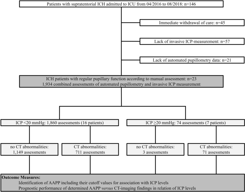
Methods: This retrospective cohort study at the neurocritical care unit of the University of Erlangen-Nuremberg (2016-2018) included 23 nontraumatic supratentorial (intracerebral hemorrhage) ICH patients without signs of abnormal pupillary function by manual assessment, i.e., absent light reflex. We assessed ICP levels by an external ventricular drain simultaneously with parameters of pupillary reactivity [i.e., maximum and minimum apertures, light reflex latency (Lat), constriction and redilation velocities (CV, DV), and percentage change of apertures (per-change)] using a portable pupillometer (NeurOptics®). Computed tomography (CT) scans were analyzed to determine lesion location, size, intraventricular hemorrhage, hydrocephalus, midline shift, and compression or absence of the basal cisterns. We performed receiver operating characteristics analysis to investigate associations of ICP levels with pupillary parameters and to determine best cutoff values for prediction of ICP elevation. After dichotomization of assessments according to ICP values (normal: < 20 mmHg, elevated: ≥ 20 mmHg), prognostic performance of the determined cutoff parameters of pupillary function versus of CT-imaging findings was analyzed by calculating sensitivity, specificity, positive and negative predictive values (logistic regression, corresponding ORs with 95% CIs).

Results: In 23 patients (11 women, median age 59.0 (51.0-69.0) years), 1,934 assessments were available for analysis. A total of 74 ICP elevations ≥ 20 mmHg occurred in seven patients. Best discriminative thresholds for ICP elevation were: CV < 0.8 mm/s (AUC 0.740), per-change < 10% (AUC 0.743), DV < 0.2 mm/s (AUC 0.703), and Lat > 0.3 s (AUC 0.616). Positive predictive value of all four parameters to indicate ICP elevation ranged between 7.2 and 8.3% only and was similarly low for CT abnormalities (9.1%). We found high negative predictive values of pupillary parameters [CV: 99.2% (95% CI 98.3-99.6), per-change: 98.7% (95% CI 97.8-99.2), DV: 98.0% (95% CI 97.0-98.7), Lat: 97.0% (95% CI 96.0-97.7)], and CT abnormalities [99.7% (95% CI 99.2-99.9)], providing evidence that both techniques adequately identified ICH patients without ICP elevation.

Conclusions: Our data suggest an association between noninvasively detected changes in pupillary reactivity and ICP levels in sedated ICH patients. Although automated pupillometry and neuroimaging seem not sufficient to noninvasively indicate ICP elevation, both techniques, however, adequately identified ICH patients without ICP elevation. This finding may facilitate routine management by saving invasive ICP monitoring or repeated CT controls in patients with specific automated pupillometry readings.

Keywords: Constriction velocity; Critical care; Intracerebral hemorrhage; Intracranial pressure; Pupillary reactivity.


Conflict of interest statement

The authors declare that they have no conflict of interest.

Figures

Fig. 1
Fig. 1
Flowchart of study participants. Overall, 146 patients with nontraumatic supratentorial ICH admitted to the ICU between April 2016 and August 2018 were screened for eligibility. After exclusion of 45 patients because of early care limitation, 57 patients because of lack of invasive ICP measurement, and 21 because of lack of automated pupillometry data, 23 ICH patients and 1,934 combined assessments of automated pupillometry and invasive ICP measurement were available for data analysis. AAPP, automatically assessed pupillary parameters; CT, computed tomography; ICH, intracerebral hemorrhage; ICP, intracranial pressure; ICU, intensive care unit
Fig. 2
Fig. 2
Automated pupillometry readings in relation to ICP levels. Constriction velocity (upper left graph), percentage change of aperture (upper right graph), dilation velocity (middle left graph), light reflex latency (middle right graph), size of aperture (lower left graph), and minimum size of aperture (lower right graph) according to intracranial pressure (ICP) values in 23 patients with nontraumatic supratentorial intracerebral hemorrhage (ICH; presented as median and interquartile range)
Fig. 3
Fig. 3
Prognostic performance of neuroimaging and automated pupillometry for identification of patients with normal versus elevated ICP levels. Hypothetical clinical scenario of 100 patients with supratentorial ICH monitored by CT imaging and automated pupillometry. Middle left and right graphs illustrate the percentage of ICH patients with (red background) and without (green background) CT abnormalities (left graph), respectively, with (red background) and without (green background) CV abnormalities (right graph). Prognostic relevance of abnormal neuroimaging and pupillometry findings for identification of ICP levels > 20 mmHg is demonstrated in the lower left and right graph. Positive predictive values of both monitoring techniques ranged less than 10% only, illustrated as red figures within the lower graphs. Gray figures visualize the high percentage of patients with CT abnormalities (left), respectively, CV abnormalities (right) despite ICP values below 20 mmHg. Prognostic relevance of both techniques for identification of ICP levels < 20 mmHg in case of absent abnormal neuroimaging and pupillometry findings is demonstrated in the upper left and right graph. Negative predictive values of CT findings (left, 99.7%) and CV (right, 99.2%) are illustrated as green figures, i.e., patients reliably identified as not at risk of ICP elevation. CT, computed tomography; CV, constriction velocity; ICP, intracranial pressure

References

    1. Hemphill JC, 3rd, et al. Guidelines for the management of spontaneous intracerebral hemorrhage: a guideline for healthcare professionals from the American Heart Association/American Stroke Association. Stroke. 2015;46(7):2032–2060. doi: 10.1161/STR.0000000000000069. - DOI - PubMed
    1. Steiner T, et al. European Stroke Organisation (ESO) guidelines for the management of spontaneous intracerebral hemorrhage. Int J Stroke. 2014;9(7):840–855. doi: 10.1111/ijs.12309. - DOI - PubMed
    1. Zhang X, et al. Invasive and noninvasive means of measuring intracranial pressure: a review. Physiol Meas. 2017;38(8):R143–R182. doi: 10.1088/1361-6579/aa7256. - DOI - PubMed
    1. Phillips SS, et al. A systematic review assessing the current state of automated pupillometry in the NeuroICU. Neurocrit Care. 2019;31(1):142–161. doi: 10.1007/s12028-018-0645-2. - DOI - PubMed
    1. Chen JW, et al. Pupillary reactivity as an early indicator of increased intracranial pressure: the introduction of the neurological Pupil index. Surg Neurol Int. 2011;2:82. doi: 10.4103/2152-7806.82248. - DOI - PMC - PubMed

Publication types