Review
. 2021 Feb;38(2):949-968.
doi: 10.1007/s12325-020-01587-5. Epub 2020 Dec 26.

Renal Tubular Acidosis and Management Strategies: A Narrative Review

Affiliations
Review

Renal Tubular Acidosis and Management Strategies: A Narrative Review

Biff F Palmer et al. Adv Ther. 2021 Feb.

Abstract

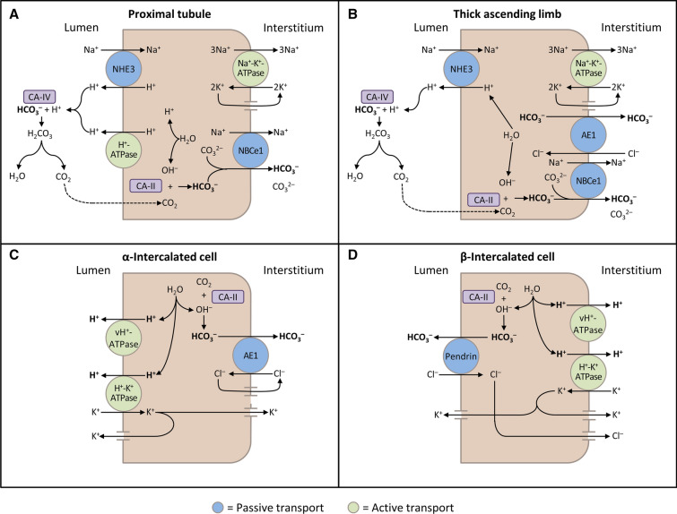
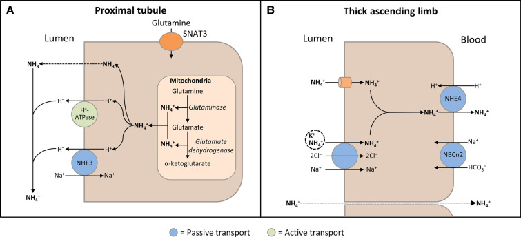
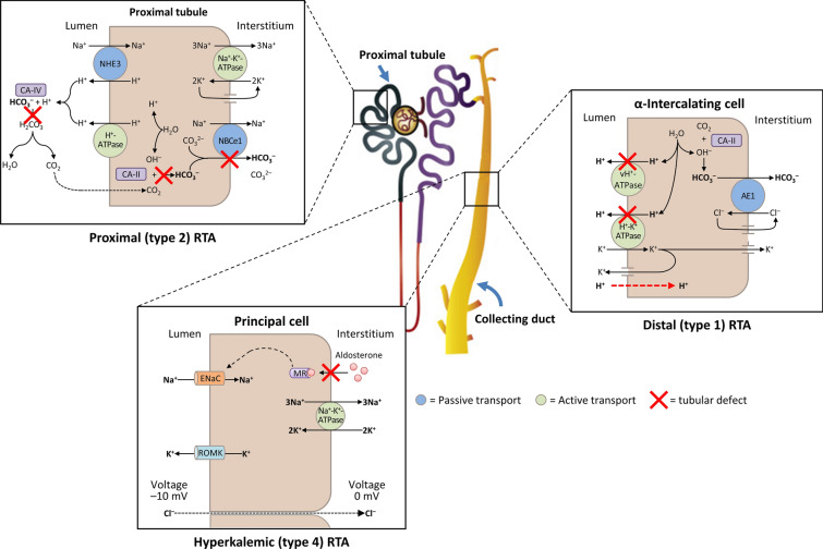
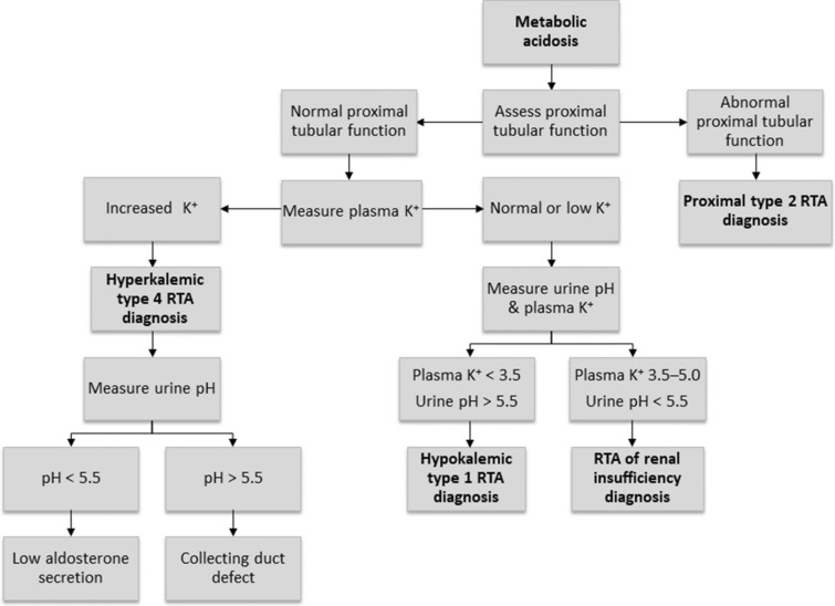
Renal tubular acidosis (RTA) occurs when the kidneys are unable to maintain normal acid-base homeostasis because of tubular defects in acid excretion or bicarbonate ion reabsorption. Using illustrative clinical cases, this review describes the main types of RTA observed in clinical practice and provides an overview of their diagnosis and treatment. The three major forms of RTA are distal RTA (type 1; characterized by impaired acid excretion), proximal RTA (type 2; caused by defects in reabsorption of filtered bicarbonate), and hyperkalemic RTA (type 4; caused by abnormal excretion of acid and potassium in the collecting duct). Type 3 RTA is a rare form of the disease with features of both distal and proximal RTA. Accurate diagnosis of RTA plays an important role in optimal patient management. The diagnosis of distal versus proximal RTA involves assessment of urinary acid and bicarbonate secretion, while in hyperkalemic RTA, selective aldosterone deficiency or resistance to its effects is confirmed after exclusion of other causes of hyperkalemia. Treatment options include alkali therapy in patients with distal or proximal RTA and lowering of serum potassium concentrations through dietary modification and potential new pharmacotherapies in patients with hyperkalemic RTA including newer potassium binders.

Keywords: Alkali therapy; Distal renal tubular acidosis; Hyperkalemic renal tubular acidosis; Normal anion gap metabolic acidosis; Potassium binders; Proximal renal tubular acidosis.


Figures

Fig. 1
Fig. 1
Schematic diagrams illustrating bicarbonate (HCO3) reabsorption and regeneration in the kidney. a HCO3 reabsorption in the proximal tubule. Hydrogen ions (H+) are secreted into the lumen via apical sodium (Na+)/H+ exchanger 3 (NHE3) and H+-ATPase transporters. Apical carbonic anhydrase (CA) IV catalyzes the reaction between H+ and HCO3, which forms H2CO3 that rapidly dissociates to water and carbon dioxide (CO2). CO2 diffuses back across the apical membrane, where CA-II catalyzes its reaction with intracellular hydroxide ions (OH) to form H+ and HCO3. HCO3 is transported across the basolateral membrane by the Na+/HCO3/CO32– cotransporter (NBCe1). b HCO3 reabsorption in the thick ascending limb. As in the proximal tubule, H+ is secreted into the lumen via NHE3, where it reacts with HCO3 to release CO2 that diffuses back across the apical membrane. HCO3 is transported across the basolateral membrane by NBCe1 and the kidney anion exchanger (AE1). c H+ secretion by α-intercalated cells in cortical collecting duct (CCD). H+ is secreted into the lumen by H+/K+-ATPase and vacuolar (v) H+-ATPase transporters on the apical membrane. Intracellular OH generated by H+ secretion reacts with CO2 via CA-II to form HCO3, which is removed by basolateral AE1. The resulting intracellular chloride (Cl) exits via conductance channels in the basolateral membrane. Luminal K+ transported into the cell via H+/K+-ATPase can exit via channels in the apical or basolateral membrane, depending on K+ balance. d HCO3 secretion by β-intercalated cells in CCD. H+-ATPase transports H+ across the basolateral membrane. Intracellular OH generated by H+ secretion reacts with CO2 via CA-II to form HCO3, which is transported into the lumen by the apical Cl/HCO3 exchanger pendrin. Intracellular Cl exits via conductance channels in the basolateral membrane (These figures were published in Comprehensive Clinical Nephrology: 5th Edition, Palmer BF, Normal acid–base balance, pp. 142–148, Copyright Elsevier (2014) [6])
Fig. 2
Fig. 2
Schematic diagrams illustrating ammonia (NH3) production and transport in the kidney. a NH3 production in the proximal tubular cells. After glutamine uptake via sodium (Na+)-coupled neutral amino acid transporter 3 (SNAT3), mitochondrial glutamine metabolism results in production of ammonium (NH4+). NH3 passively diffuses across apical membrane and hydrogen (H+) is transported via apical Na+/H+ exchanger 3 (NHE3) and H+-ATPase; NH3 and H+ combine in the lumen to form NH4+. b NH3 transport in the thick ascending limb. Lumen-positive voltage drives passive paracellular transport of NH4+ from the lumen into the blood. By substituting for potassium (K+), NH4+ is also transported into the cell via the Na+/K+/2Cl transporter and the apical membrane K+ channel (ROMK). The basolateral Na+/bicarbonate (HCO3) cotransporter (NBCn2) may play a role in maintaining cellular pH. NH4+ crosses the basolateral membrane into the blood via NHE4 (Adapted with permission from Palmer 2014 [6])
Fig. 3
Fig. 3
A schematic diagram illustrating the underlying kidney tubule defects causing the different types of renal tubular acidosis (RTA). Distal (type 1) RTA is caused by either impaired hydrogen (H+) secretion by vacuolar (v) H+-ATPase or H+/K+-ATPase or increased H+ permeability of luminal membrane by α-intercalated cells of the collecting duct, which leads to a reduction in net H+ secretion. Proximal (type 2) RTA is caused by defects in bicarbonate (HCO3) reabsorption in the proximal tubule, due to either impaired HCO3 transport across the basolateral membrane or inhibition of carbonic anhydrase (CA) activity. Hyperkalemic (type 4) RTA is caused by aldosterone deficiency or resistance, which leads to reduced Na+ (sodium) reabsorption by principal cells of the collecting duct and decreased transepithelial voltage, leading to diminished H+ secretion by α-intercalated cells and K+ secretion by principal cells. AE1 kidney anion exchanger, ENaC epithelial Na+ channel, MR mineralocorticoid receptor, NHE3 Na+/H+ exchanger 3, ROMK apical membrane K+ channel
Fig. 4
Fig. 4
A suggested algorithm for diagnosing renal tubular acidosis. Reprinted by permission of Edizioni Minerva Medica from Minerva Endocrinologica 2019 December; 44(4):363–77 [7]

Similar articles

Cited by

References

    1. Hamm LL, Nakhoul N, Hering-Smith KS. Acid-base homeostasis. Clin J Am Soc Nephrol. 2015;10:2232–2242. doi: 10.2215/CJN.07400715. - DOI - PMC - PubMed
    1. Scialla JJ, Asplin J, Dobre M, et al. Higher net acid excretion is associated with a lower risk of kidney disease progression in patients with diabetes. Kidney Int. 2017;91:204–215. doi: 10.1016/j.kint.2016.09.012. - DOI - PMC - PubMed
    1. Halperin ML, Jungas RL. Metabolic production and renal disposal of hydrogen ions. Kidney Int. 1983;24:709–713. doi: 10.1038/ki.1983.217. - DOI - PubMed
    1. Hamilton PK, Morgan NA, Connolly GM, Maxwell AP. Understanding acid-base disorders. Ulster Med J. 2017;86:161–166. - PMC - PubMed
    1. Soleimani M, Rastegar A. Pathophysiology of renal tubular acidosis: core curriculum 2016. Am J Kidney Dis. 2016;68:488–498. doi: 10.1053/j.ajkd.2016.03.422. - DOI - PubMed

Publication types