. 2020 Dec;120(12):1700-1715.
doi: 10.1055/s-0040-1721319. Epub 2020 Dec 23.

Heparin Inhibits Cellular Invasion by SARS-CoV-2: Structural Dependence of the Interaction of the Spike S1 Receptor-Binding Domain with Heparin

Affiliations

Heparin Inhibits Cellular Invasion by SARS-CoV-2: Structural Dependence of the Interaction of the Spike S1 Receptor-Binding Domain with Heparin

Courtney J Mycroft-West et al. Thromb Haemost. 2020 Dec.

Abstract

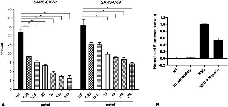
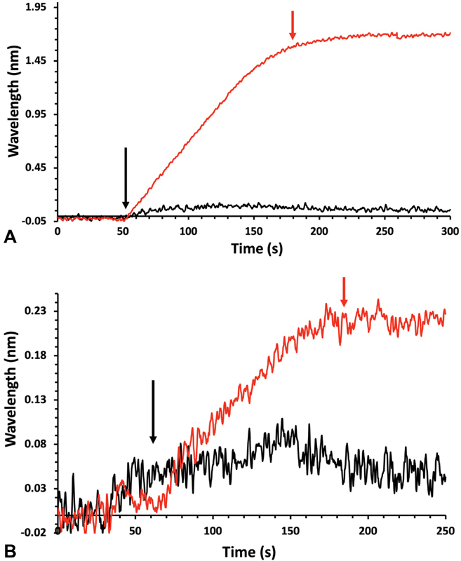
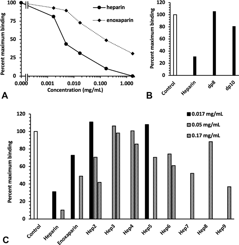
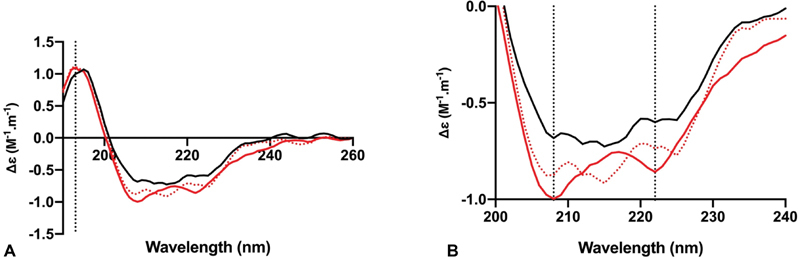
The dependence of development and homeostasis in animals on the interaction of hundreds of extracellular regulatory proteins with the peri- and extracellular glycosaminoglycan heparan sulfate (HS) is exploited by many microbial pathogens as a means of adherence and invasion. Heparin, a widely used anticoagulant drug, is structurally similar to HS and is a common experimental proxy. Exogenous heparin prevents infection by a range of viruses, including S-associated coronavirus isolate HSR1. Here, we show that heparin inhibits severe acute respiratory syndrome coronavirus-2 (SARS-CoV-2) invasion of Vero cells by up to 80% at doses achievable through prophylaxis and, particularly relevant, within the range deliverable by nebulisation. Surface plasmon resonance and circular dichroism spectroscopy demonstrate that heparin and enoxaparin, a low-molecular-weight heparin which is a clinical anticoagulant, bind and induce a conformational change in the spike (S1) protein receptor-binding domain (S1 RBD) of SARS-CoV-2. A library of heparin derivatives and size-defined fragments were used to probe the structural basis of this interaction. Binding to the RBD is more strongly dependent on the presence of 2-O or 6-O sulfate groups than on N-sulfation and a hexasaccharide is the minimum size required for secondary structural changes to be induced in the RBD. It is likely that inhibition of viral infection arises from an overlap between the binding sites of heparin/HS on S1 RBD and that of the angiotensin-converting enzyme 2. The results suggest a route for the rapid development of a first-line therapeutic by repurposing heparin and its derivatives as antiviral agents against SARS-CoV-2 and other members of the Coronaviridae.


Conflict of interest statement

None declared.

Figures

Fig. 1
Fig. 1
The heparin-mediated inhibition of SARS-CoV-2 viral invasion of Vero cells. ( A ) The effect of unfractionated porcine mucosal heparin added 1 hour before the infection of Vero cells with 50 PFU of SARS-CoV-2 or SARS-CoV. The results are expressed as the number of PFU per well and represent the mean ± SD of quadruplicate cultures. The p -value was calculated using the Mann–Whitney U test, P  ≤ 0.05; ∗∗ P  ≤ 0.01; ∗∗∗ P  ≤ 0.001. ( B ) The effect of unfractionated porcine mucosal heparin (100 μg mL −1 ) on RBD binding to Vero cells. Nil represents no treatment; no secondary represents no secondary antibody control; au, arbitrary units of fluorescence. PFU, plaque-forming unit; RBD, receptor-binding domain; SD, standard deviation.
Fig. 2
Fig. 2
Interaction of FGF2 and 800 nM SARS-CoV-2 S1 RBD with immobilised heparin. Reducing end biotinylated heparin was immobilised on a streptavidin-functionalised P4SPR sensor surface [no biotin-heparin (−) control]. PBS running buffer flow rate was 500 µL min −1 . The data for the three sensing channels are reported as an average response (−). The start of protein injections is indicated by black arrows and the return of the surface to the running buffer (PBST) by red arrows . ( A ) Injection of 100 nM FGF2. ( B ) Injection of 800 nM SARS-CoV-2 S1 RBD protein. PBS, phosphate buffered saline; PBST, phosphate buffered saline with Tween-20; RBD, receptor-binding domain.
Fig. 3
Fig. 3
Competition for SARS-CoV-2 S1 RBD binding to immobilised heparin with model heparin-derived oligo- and polysaccharides. 800 nM SARS-CoV-2 S1 RBD was injected onto the surface in the presence or absence of the indicated concentration of heparin-derived oligo- and polysaccharides. Since the SARS-CoV-2 S1 RBD does not dissociate appreciably when the system returns to PBST, this is when the response was measured, to avoid any confounding effects of differences of refractive index between samples. A control measurement (800 nM SARS-CoV-2 S1 RBD alone) was performed before each competition and used to calculate the percentage of maximum binding. This ensured that small changes over time in the responsiveness of the surface did not confound the analysis. ( A ) Competition for 800 nM SARS-CoV-2 S1 RBD binding to immobilised heparin by heparin and by enoxaparin. ( B ) Competition for 800 nM SARS-CoV-2 S1 RBD binding to immobilised heparin by 0.17 mg mL −1 heparin-derived dp 8 and dp 10, with the corresponding value for heparin from panel (A) shown to aid comparison. ( C ) Competition for 800 nM SARS-CoV-2 S1 RBD binding to immobilised heparin by a panel of orthogonally chemically desulfated heparins ( Table 1 ). PBST, phosphate buffered saline with Tween-20; RBD, receptor-binding domain.
Fig. 4
Fig. 4
The conformational change of the SARS-CoV-2 S1 RBD observed in the presence of heparin by CD spectroscopy. ( A ) Circular dichroism spectra (190–260 nm) of SARS-CoV-2 S1 RBD alone ( black solid line ) and with heparin ( red solid line ) in PBS, pH 7.4. The red, dotted line represents the sum of the two individual spectra and the fact that this is distinct from the spectrum of the RBD with heparin ( red solid line ) indicates that a conformational change and, therefore, binding have occurred. The dotted vertical line indicates 193 nm. ( B ) Details of the same spectra expanded between 200 and 240 nm. Vertical dotted lines indicate 222 and 208 nm. PBS, phosphate buffered saline; RBD, receptor-binding domain.
Fig. 5
Fig. 5
The conformational change of the SARS-CoV-2 S1 RBD observed in the presence of a chemically modified heparin derivative by CD spectroscopy. ( A ) Circular dichroism spectra (190–260 nm) of SARS-CoV-2 S1 RBD alone ( black solid line ) with heparin ( red solid line ) and with a chemically modified derivative, heparin 5 ( Table 1 ), with the predominant repeating disaccharide structure; –IdoA2OH-GlcNAc6S–( blue solid line ) in PBS, pH 7.4. The vertical dotted line indicates 193 nm ( B ). The same spectra expanded between 200 and 240 nm. Vertical dotted lines indicate 222 and 208 nm. ( C ) Secondary structure content analysed using BeStSel (20) for SARS-Cov-2 S1 RBD [analysis using BeStSel was performed on smoothed CD data from (A) between 190 and 260 nm]. PBS, phosphate buffered saline; RBD, receptor-binding domain.
Fig. 6
Fig. 6
The conformational change of the SARS-CoV-2 S1 RBD observed in the presence of size-defined heparin oligosaccharides by CD spectroscopy. Circular dichroism spectra between 200 and 240 nm of SARS CoV-2 S1 RBD in PBS, pH 7.4, alone ( black solid line ), with heparin ( red solid line ), and PMH-derived, size-defined oligosaccharides ( blue solid line ): ( A ) Tetrasaccharide, ( B ) hexasaccharide, ( C ) octasaccharide, and ( D ) decasaccharide. Vertical dotted lines indicate 222 and 208 nm. PBS, phosphate buffered saline; RBD, receptor-binding domain.
Fig. 7
Fig. 7
SARS-CoV-2 S1 RBD protein model. Basic amino acids that are solvent accessible on the surface are indicated ( dark blue ); these form extensive patches. Sequences with the highest normalised count ( Tables 2 and 3 ) are highlighted. R346 is also shown as it indicates a potential heparin-binding gain of function mutation (T346R) from the Bat-RaTG13. RBD, receptor-binding domain.
Fig. 8
Fig. 8
Snapshots taken from MD simulations of SARS-CoV-RBD in the presence of either a heparin octasaccharide (A) or a heparin hexasaccharide ( B ). The heparin oligosaccharides are shown as sticks whereas amino acids of the RBD are shown as spheres. The residues are coloured as per elements. Hydrogen atoms are not shown for clarity. The regions subjected to conformational changes in the protein during the simulations are highlighted in yellow ribbon. MD, molecular dynamics; RBD, receptor-binding domain.

References

    1. Woznica A, Gerdt J P, Hulett R E, Clardy J, King N. Mating in the closest living relatives of animals is induced by a bacterial chondroitinase. Cell. 2017;170(06):1.175E14–1.183E14. - PMC - PubMed
    1. Ori A, Wilkinson M C, Fernig D G. A systems biology approach for the investigation of the heparin/heparan sulfate interactome. J Biol Chem. 2011;286(22):19892–19904. - PMC - PubMed
    1. Ireland E V, Woznica A, King N. Synergistic cues from diverse bacteria enhance multicellular development in a choanoflagellate. Appl Environ Microbiol. 2020;86(11):e02920-19. - PMC - PubMed
    1. Ori A, Wilkinson M C, Fernig D G. The heparanome and regulation of cell function: structures, functions and challenges. Front Biosci. 2008;13:4309–4338. - PubMed
    1. Nunes Q M, Su D, Brownridge P J. The heparin-binding proteome in normal pancreas and murine experimental acute pancreatitis. PLoS One. 2019;14(06):e0217633. - PMC - PubMed

MeSH terms