. 2021 Jun;529(9):2362-2375.
doi: 10.1002/cne.25100. Epub 2021 Jan 7.

Defining vitamin D receptor expression in the brain using a novel VDRCre mouse

Affiliations

Defining vitamin D receptor expression in the brain using a novel VDRCre mouse

Hailan Liu et al. J Comp Neurol. 2021 Jun.

Abstract

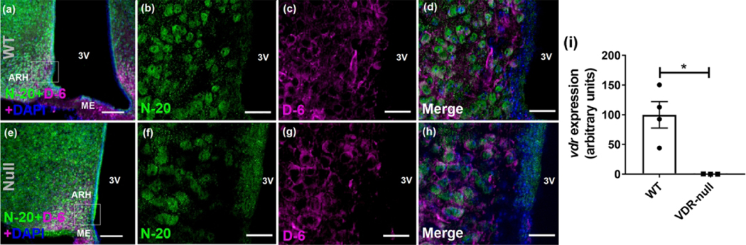
Vitamin D action has been linked to several diseases regulated by the brain including obesity, diabetes, autism, and Parkinson's. However, the location of the vitamin D receptor (VDR) in the brain is not clear due to conflicting reports. We found that two antibodies previously published as specific in peripheral tissues are not specific in the brain. We thus created a new knockin mouse with cre recombinase expression under the control of the endogenous VDR promoter (VDRCre ). We demonstrated that the cre activity in the VDRCre mouse brain (as reported by a cre-dependent tdTomato expression) is highly overlapping with endogenous VDR mRNAs. These VDR-expressing cells were enriched in multiple brain regions including the cortex, amygdala, caudate putamen, and hypothalamus among others. In the hypothalamus, VDR partially colocalized with vasopressin, oxytocin, estrogen receptor-α, and β-endorphin to various degrees. We further functionally validated our model by demonstrating that the endogenous VDR agonist 1,25-dihydroxyvitamin D activated all tested tdTomato+ neurons in the paraventricular hypothalamus but had no effect on neurons without tdTomato fluorescence. Thus, we have generated a new mouse tool that allows us to visualize VDR-expressing cells and to characterize their functions.

Keywords: RRID:AB_141637; RRID:AB_2157629; RRID:AB_2314007; RRID:AB_2715552; RRID:AB_2832252; RRID:AB_310305; RRID:AB_628040; RRID:AB_632069; brain; immunohistochemistry; mutant mouse strain; vitamin D receptor.


Figures

Fig. 1.
Fig. 1.
Dual immunohistochemical staining for the VDR using different antibodies in the hypothalamus. a-d, Low (a) and high (b-d) magnification images of antibody staining in wild type animals with N-20 (b), D-6 (c), and merged image (d). e-h, Low (e) and high (f-h) magnification images of antibody staining in VDR-null animals with N-20 (f), D-6 (g), and merged image (h). (i) vdr gene expression in whole hypothalamic samples of VDR-null mice normalized to L32 in all samples. For all images, N-20 is green, D-6 is magenta, and DAPI is blue. Box area delineates area of higher magnification. Scale bars: 100 μm in widefield view (a, e) and 25 μm in higher magnification (b-d, f-h). 3V, third ventricle; ARH, arcuate hypothalamus; ME, median eminence; WT, wild type. * p < 0.05.
Fig. 2.
Fig. 2.
Development and phenotype of VDRCre mice. (a) Schematic construction of the VDRCre transgenic mouse. Cre recombinase was targeted to the C-terminal of the VDR gene with a GGGGS linker, V5 tag and P2A sequence. Primer sequences A-E refer to sequences listed in Table 2. (b) Body weight comparison of VDRCre;Ai14 vs. control mice (n = 5–8/gp). (c) Intraperitoneal glucose tolerance test (1.5 g/kg dextrose) in same mice. (d) vdr expression in duodenum, kidney, and liver in VDRCre;Ai14 mice is similar to wild type controls (n=3–4 mice/tissue/group; samples normalized to tissue-specific housekeeping genes and then to average control duodenum expression). (e) tdTomato expression is significantly higher in duodenum and kidney of VDRCre;Ai14 mice, with no expression in liver (n=3–4 mice/tissue/group; samples normalized as in figure 2d). (f) vdr expression in hippocampal tdTomato+ neurons is significantly higher than in tdTomato- mice (n=8 neurons from one mouse/group; normalized to L32). (g) Absolute counts of tdTomato+ cells of VDRCre; Ai14 mice in areas of the whole brain (numbers are averages of 4–8 sections/mouse in 3 different mice per sex). * P < 0.05
Fig. 3.
Fig. 3.
Expression of tdTomato mRNAs and colocalization with VDR mRNAs in VDRCre male mice (part 1). Pictures of different brain areas are shown for tdTomato mRNAs (TD; magenta) and VDR mRNAs (VDR; green). Merged images show co-localized cells in white. Boxed area in merged image is enlarged in the adjacent image. BNST, bed nucleus of the stria terminalis; CPu, caudate putamen; Rt, reticular thalamic nucleus; LV, lateral ventricle. Scale bar = 100 μm for all except enlarged images (d, h, l, p, t) where scale bar = 25 μm.
Fig. 4.
Fig. 4.
Expression of tdTomato mRNAs and colocalization with VDR mRNAs in VDRCre male mice (part 2). Pictures of different brain areas are shown for tdTomato mRNAs (TD; magenta) and VDR mRNAs (VDR; green). Merged images show co-localized cells in white. Boxed area in merged image is enlarged in the adjacent image. 3V, third ventricle; Aq, aqueduct; ARH, arcuate hypothalamus; DMH, dorsomedial hypothalamus, DRN, dorsal Raphe nucleus; PVH, paraventricular hypothalamus, VMH, ventromedial hypothalamus. Scale bar = 100 μm for all except enlarged images (d, h, l, p, t) where scale bar = 25 μm.
Fig. 5.
Fig. 5.
Co-expression of key cell markers with VDR expression in the hypothalamus of male mice. Expression of tdTomato (Tom; magenta) and cell markers (green) for vasopressin (AVP; b), oxytocin (OXY;e), ERα (h, k), β-endorphin (n). Arrows denote cells with colocalization of tdTomato and GFP fluorescence. 3V, third ventricle; ARH, arcuate hypothalamus; PVH, paraventricular hypothalamus. Scale bar = 50 μm.
Fig. 6.
Fig. 6.
Effects of VDR agonist on neural activities of tdTomato+ and tdTomato cells in the PVH. a. Representative current clamp traces in response to vitamin D (1,25D3, 5μM, puff) in tdTomato+ and tdTomato cells and to vehicle in tdTomato+ cells. b, c. Summary of resting membrane potential (b) and firing frequency (c) before and after vitamin D puff in female mice. d, e. Summary of resting membrane potential (d) and firing frequency (e) in tdTomato+ cells before and after vehicle puff in female mice. f, g. Summary of resting membrane potential (f) and firing frequency (g) before and after vitamin D puff in male mice. h, i. Summary of resting membrane potential (h) and firing frequency (i) in tdTomato+ cells before and after vehicle puff in male mice. j. Representative current clamp trace in response to vitamin D (5μM, puff) in tdTomato+ cells in the presence of 1 μM TTX, 30 μM CNQX, 30 μM D-AP5, and 50 μM bicuculline. k, l. Summary data of resting membrane potential before and after vitamin D puff in the presence of 1 μM TTX, 30 μM CNQX, 30 μM D-AP5, and 50 μM bicuculline. Data are presented for each cell collected from 2 male and 2 female mice. ***, P < 0.001 in paired t-tests.

Similar articles

Cited by

References

    1. Bassett AR, Tibbit C, Ponting CP, & Liu J-L (2013). Highly Efficient Targeted Mutagenesis of Drosophila with the CRISPR/Cas9 System. Cell Reports, 4(1), 220–228. 10.1016/j.celrep.2013.06.020 - DOI - PMC - PubMed
    1. Biswas P, & Zanello LP (2009). 1alpha,25(OH)(2) vitamin D(3) induction of ATP secretion in osteoblasts. Journal of Bone and Mineral Research : The Official Journal of the American Society for Bone and Mineral Research, 24(8), 1450–1460. 10.1359/jbmr.090306 - DOI - PMC - PubMed
    1. Cao X, Xu P, Oyola MG, Xia Y, Yan X, Saito K, Zou F, Wang C, Yang Y, Jr AH, Yan C, Ding H, Zhu L, Yu L, Yang B, Feng Y, Clegg DJ, Khan S, Dimarchi R, … Xu Y. (2014). Estrogens stimulate serotonin neurons to inhibit binge-like eating in mice. 10.1172/JCI74726 - DOI - PMC - PubMed
    1. Choe HK, Reed MD, Benavidez N, Montgomery D, Soares N, Yim YS, & Choi GB (2015). Oxytocin Mediates Entrainment of Sensory Stimuli to Social Cues of Opposing Valence. Neuron, 87(1), 152–163. 10.1016/j.neuron.2015.06.022 - DOI - PMC - PubMed
    1. Cui X, Pelekanos M, Liu P-Y, Burne THJ, McGrath JJ, & Eyles DW (2013). The vitamin D receptor in dopamine neurons; its presence in human substantia nigra and its ontogenesis in rat midbrain. Neuroscience, 236, 77–87. 10.1016/j.neuroscience.2013.01.035 - DOI - PubMed

Publication types

LinkOut - more resources