. 2021 Feb;25(4):2190-2202.
doi: 10.1111/jcmm.16204. Epub 2020 Dec 25.

The anti-angiogenesis role of FBXW7 in diabetic retinopathy by facilitating the ubiquitination degradation of c-Myc to orchestrate the HDAC2

Affiliations

The anti-angiogenesis role of FBXW7 in diabetic retinopathy by facilitating the ubiquitination degradation of c-Myc to orchestrate the HDAC2

Lihua Hu et al. J Cell Mol Med. 2021 Feb.

Abstract

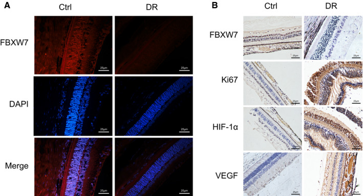
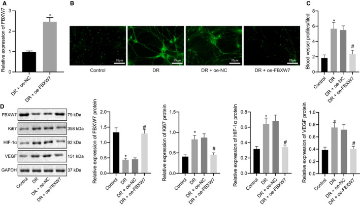
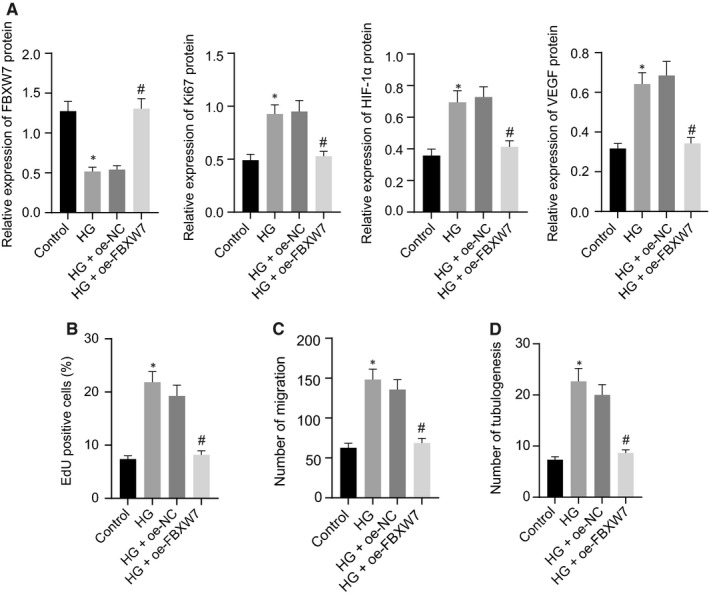
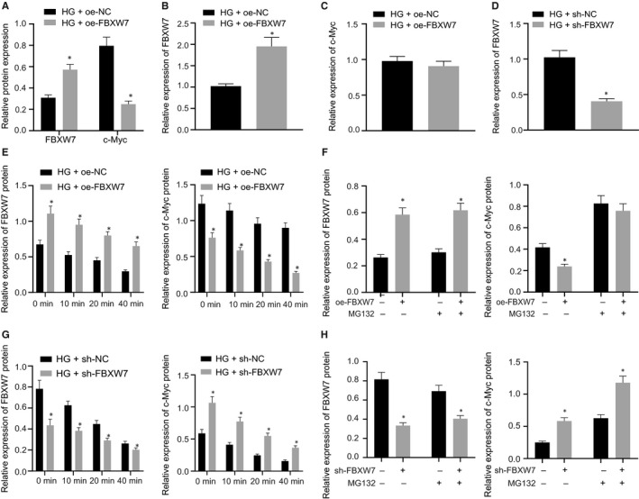
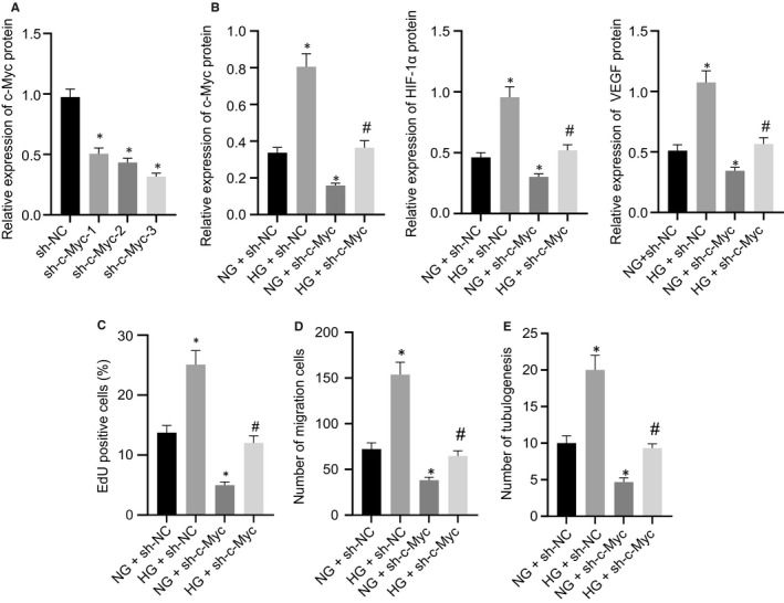
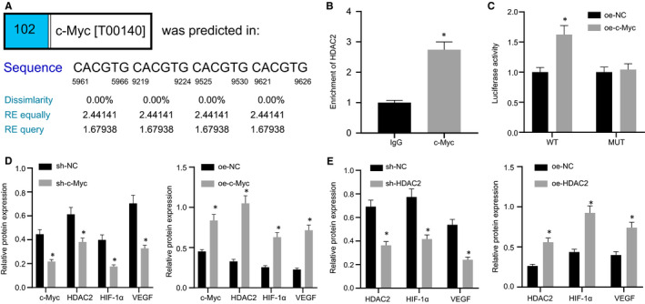
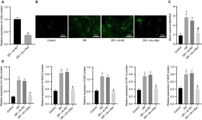
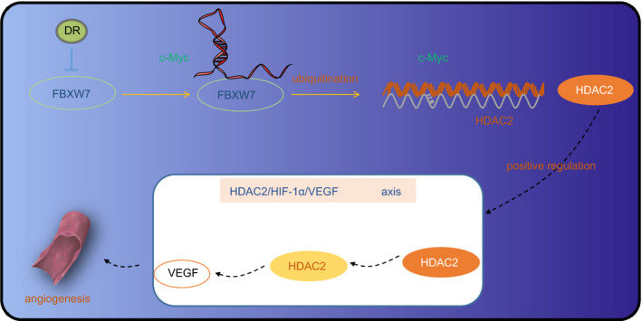
Diabetic retinopathy (DR) is the most prevalently occurring microvascular complication in diabetic patients that triggers severe visual impairments. The anti-angiogenesis role of FBXW7 has been identified in breast cancer. Therefore, this study intends to decipher the mechanism of FBXW7 in angiogenesis of DR. DR model was induced on mice using high-glucose (HG) and high-fat diet, and retinal microvascular endothelial cells (RMECs) isolated from normal mice were induced with HG, followed by evaluation of FBXW7, Ki67, HIF-1α and VEGF expression by immunofluorescence, immunohistochemistry or Western blot analysis. After gain- and loss-of-function assays in normal and DR mice, angiogenesis was assessed by CD31 fluorescence staining and Western blot analysis. After ectopic expression and silencing experiments in HG-induced RMECs, RMEC proliferation, migration and angiogenesis were, respectively, determined by EdU, Transwell and in vitro angiogenesis assays. The impact of FBXW7 on the ubiquitination of c-Myc was studied by cycloheximide chase assay and proteasome inhibition, and the binding of c-Myc to HDAC2 promoter by dual-luciferase reporter gene experiment. DR mice and HG-induced RMECs possessed down-regulated FBXW7 and up-regulated Ki67, HIF-1α and VEGF. Silencing FBXW7 enhanced angiogenesis in normal mouse retinal tissue, but overexpressing FBXW7 or silencing c-Myc diminished angiogenesis in DR mouse retinal tissue. Overexpressing FBXW7 or silencing c-Myc depressed proliferation, migration and angiogenesis in HG-induced RMECs. FBXW7 induced c-Myc ubiquitination degradation, and c-Myc augmented HDAC2 expression by binding to HDAC2 promoter. Conclusively, our data provided a novel sight of anti-angiogenesis role of FBXW7 in DR by modulating the c-Myc/HDAC2 axis.

Keywords: Angiogenesis; Diabetic retinopathy; FBXW7; HDAC2; HIF-1α; VEGF; c-Myc.


Conflict of interest statement

All authors declare that they have no conflicts of interests.

Figures

Figure 1
Figure 1
FBXW7 is highly expression but Ki67, HIF‐1α and VEGF are poorly expressed in the retinal tissue of DR mice. (A) Immunofluorescence detection of FBXW7 expression in retinal tissues of control mice (n = 6) and DR mice (n = 6) (scale: 25 μm). (B) Immunohistochemical detection of FBXW7, Ki67, HIF‐1α and VEGF expression in retinal tissues of control mice (n = 6) and DR mice (n = 6) (scale: 25 μm)
Figure 2
Figure 2
Silencing FBXW7 in normal mouse retina promotes angiogenesis. (A) RT‐qPCR identification of the silencing efficiency of FBXW7 in RMECs. * P < .05 vs. sh‐NC‐treated RMECs. Normal diet‐fed mice were treated with sh‐NC or sh‐FBXW7. (B) RT‐qPCR detection of the silencing efficiency of sh‐FBXW7 in the retina of normal diet‐fed mice. (C) Representative image of CD31 fluorescence staining of intraretinal angiogenesis in the retina of normal diet‐fed mice (Arrows indicated blood vessel profiles [BVPs], scale: 25 μm.). (D) Quantitative results of panel C. E, Western blot analysis of FBXW7, HIF‐1α, Ki67 and VEGF expression in the retina of normal diet‐fed mice. * P < .05 vs. normal diet‐fed mice treated with sh‐NC. The measurement data were expressed as mean ± standard deviation. The unpaired t test was used for comparison between two groups. The cell experiments were repeated 3 times. n = 6 mice/group
Figure 3
Figure 3
Overexpression of FBXW7 inhibits intraretinal angiogenesis in DR mice. (A) RT‐qPCR detection of the overexpression efficiency of FBXW7 in mice. * P < .05 vs. oe‐NC‐treated DR mice. Mice were fed with normal diet as control, whereas DR mice were untreated, or treated with oe‐NC or oe‐FBXW7. (B) Representative image of CD31 fluorescence staining of intraretinal angiogenesis (Arrows indicate blood vessel profiles [BVPs], scale: 25 μm.). (C) Quantitative results of panel B. (D) Western blot analysis of FBXW7, HIF‐1α, Ki67 and VEGF expression in mouse retina. * P < .05 vs. normal diet‐fed mice; # P < .05 vs. DR mice treated with oe‐NC. The measurement data were expressed as mean ± standard deviation. The unpaired t test was used for comparison between two groups, and Tukey's test‐corrected one‐way ANOVA was used for comparison among multiple groups. n = 6 mice/group
Figure 4
Figure 4
Overexpression of FBXW7 reduces HG‐induced proliferation, migration and angiogenesis of RMECs. NG‐treated RMECs were used as controls, whereas HG‐treated RMECs were uninfected, or infected with oe‐NC or oe‐FBXW7 lentivirus. (A) Western blot analysis of FBXW7, Ki67, HIF‐1α and VEGF expression in RMECs. (B) EdU assay of RMEC proliferation. (C) Transwell assay of RMEC migration. (D) Angiogenesis assay in vitro to identify the tube formation of RMECs. * P < .05 vs. NG‐treated RMECs; # P < .05 vs. HG‐treated RMECs transfected with oe‐NC. The measurement data were expressed as mean ± standard deviation. Tukey's test‐corrected one‐way ANOVA was used for comparison among multiple groups. The experiments were repeated 3 times
Figure 5
Figure 5
FBXW7 promotes c‐Myc ubiquitination degradation in HG‐induced RMECs. (A) Western blot analysis of FBXW7 and c‐Myc protein expression in HG‐induced RMECs after overexpression of FBXW7. (B) RT‐qPCR detection of FBXW7 mRNA level in HG‐induced RMECs after overexpression of FBXW7. (C) RT‐qPCR detection of the mRNA level of c‐Myc in HG‐induced RMECs after overexpression of FBXW7. *P < .05 vs. HG‐induced RMECs transfected with oe‐NC. (D) RT‐qPCR detection of the silencing efficiency of FBXW7 in HG‐induced RMECs. *P < .05 vs. HG‐induced RMECs transfected with sh‐NC. (E) Western blot analysis of FBXW7 and c‐Myc expression in HG‐induced RMECs after FBXW7 overexpression and different times of CHX (100 μg/mL) treatment. (F) Western blot analysis of FBXW7 and c‐Myc expression in HG‐induced RMECs after infection with oe‐NC or oe‐FBXW7 lentivirus and treatment with or without MG132 for 8 h. (G) Western blot analysis of the expression of FBXW7 and c‐Myc in HG‐induced RMECs after infection with sh‐NC or sh‐FBXW7 lentivirus and different times of CHX (100 μg/mL) treatment. (H) Western blot analysis of the expression of FBXW7 and c‐Myc in HG‐induced RMECs after infection with sh‐NC or sh‐FBXW7 lentivirus and treatment with or without MG132 for 8 h. * P < .05 vs. control treatment. The measurement data were expressed as mean ± standard deviation. The unpaired t test was used for comparison between two groups. The experiments were repeated 3 times
Figure 6
Figure 6
c‐Myc silencing rescues the increased angiogenesis of RMECs caused by HG. (A) Silencing efficiency of sh‐c‐Myc in RMECs. * P < .05 vs. RMECs infected with sh‐NC lentivirus. NG‐induced RMECs were infected with sh‐NC or sh‐c‐Myc lentivirus, whereas HG‐induced RMECs were infected with sh‐NC or sh‐c‐Myc lentivirus. (B) Western blot analysis of HIF‐1α and VEGF expression in RMECs. (C) EdU assay of RMEC proliferation. (D) Transwell assay to detect RMEC migration. (E) Angiogenesis assays in vitro to detect the tube formation of RMECs. * P < .05 vs. NG‐induced RMECs treated with sh‐NC; # P < .05 vs. HG‐induced RMECs treated with sh‐NC. The measurement data were expressed as mean ± standard deviation. Tukey's test‐corrected one‐way ANOVA was adopted for comparison among multiple groups. The experiments were repeated 3 times
Figure 7
Figure 7
c‐Myc elevates HDAC2 expression by binding to the HDAC2 promoter. (A) Prediction of the binding site of c‐Myc to the HDAC2 promoter region using PROMO. (B) ChIP experiment to detect the binding of c‐Myc and HDAC2 promoter in RMECs. (C) Dual‐luciferase reporter assay to verify the binding of c‐Myc to HDAC2 promoter. (D) Western blot analysis of c‐Myc, HDAC2, HIF‐1α and VEGF expression in HG‐induced RMECs after overexpressing or silencing c‐Myc. (E) Western blot analysis the expression of HDAC2, HIF‐1α and VEGF in HG‐induced RMECs after overexpressing or silencing HDAC2. * P < .05 vs. treatment with IgG, sh‐NC or oe‐NC. The measurement data were expressed as mean ± standard deviation. The unpaired t test was used for comparison between two groups. The experiments were repeated 3 times
Figure 8
Figure 8
FBXW7‐mediated ubiquitination degradation of c‐Myc regulates the HDAC2/HIF‐1α/VEGF axis to inhibit angiogenesis in DR mice. (A) RT‐qPCR detection of the silencing efficiency of c‐Myc in the retina of DR mice. * P < .05 vs. sh‐NC‐treated DR mice. Mice were fed with normal diet as control, whereas DR mice were untreated, or treated with sh‐NC or sh‐c‐Myc. (B) Representative image of CD31 fluorescence staining of intraretinal angiogenesis (Arrows indicate blood vessel profiles [BVPs], scale: 25 μm.). (C) Quantitative results of panel B. (D) Western blot analysis of c‐Myc, HDAC2, Ki67, HIF‐1α and VEGF expression in the mouse retina. * P < .05 vs. normal diet‐fed mice; # P < .05 vs. DR mice treated with sh‐NC. The measurement data were expressed as mean ± standard deviation. The unpaired t test was used for comparison between two groups, and Tukey's test‐corrected one‐way ANOVA was used for comparison among multiple groups. n = 6 mice/group
Figure 9
Figure 9
FBXW7 inhibits angiogenesis in DR via HDAC2/HIF‐1α/VEGF axis by promoting the ubiquitination degradation of c‐Myc

References

    1. Potilinski MC, Lorenc V, Perisset S, et al. Mechanisms behind retinal ganglion cell loss in diabetes and therapeutic approach. Int J Mol Sci. 2020;21. - PMC - PubMed
    1. Vujosevic S, Aldington SJ, Silva P, et al. Screening for diabetic retinopathy: new perspectives and challenges. Lancet Diabetes Endocrinol. 2020;8:337‐347. - PubMed
    1. Sabanayagam C, Banu R, Chee ML, et al. Incidence and progression of diabetic retinopathy: a systematic review. Lancet Diabetes Endocrinol. 2019;7:140‐149. - PubMed
    1. Stitt AW, Curtis TM, Chen M, et al. The progress in understanding and treatment of diabetic retinopathy. Prog Retin Eye Res. 2016;51:156‐186. - PubMed
    1. Lee VK, Hosking BM, Holeniewska J, et al. BTBR ob/ob mouse model of type 2 diabetes exhibits early loss of retinal function and retinal inflammation followed by late vascular changes. Diabetologia. 2018;61:2422‐2432. - PMC - PubMed

MeSH terms