Review
. 2021 Apr;34(2):70-79.
doi: 10.1177/1971400920983566. Epub 2020 Dec 28.

Atypia of undetermined significance/follicular lesions of undetermined significance: What radiologists need to know

Affiliations
Review

Atypia of undetermined significance/follicular lesions of undetermined significance: What radiologists need to know

Johnny Ling et al. Neuroradiol J. 2021 Apr.

Abstract

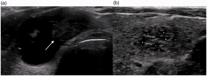
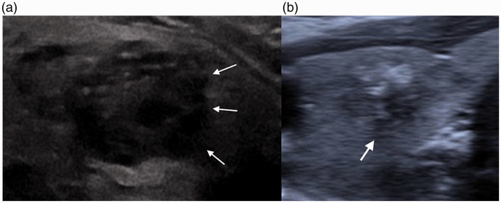
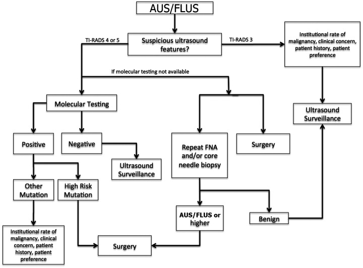
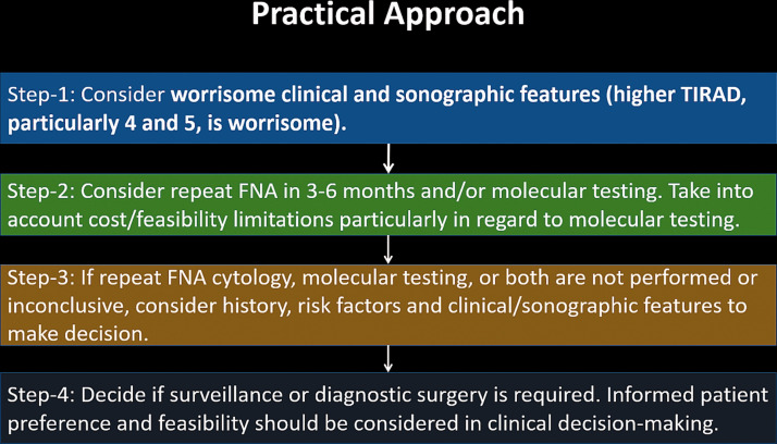
Atypia of undetermined significance/follicular lesions of undetermined significance (AUS/FLUS) refers to an intermediate histologic category of thyroid nodules in The Bethesda System for Reporting Thyroid Cytopathology. Although the risk of malignancy in this category was originally cited as 5-15%, recent literature has suggested higher rates of related malignancy ranging from 38% to 55%. Malignant nodules warrant surgery with total thyroidectomy or thyroid lobectomy, whereas benign nodules can be observed or followed with serial ultrasounds (US) based on their imaging characteristics. The management of nodules with a cytopathologic diagnosis of AUS/FLUS can be difficult because theses nodules lie between the extremes of benign and malignant. The management options for such nodules include observation, repeat fine-needle aspiration, and surgery. The use of molecular genetics, the identification of suspicious US characteristics, and the recognition of additional clinical factors are all important in the development of an appropriate, tailored management approach. Institutional factors also play a crucial role.

Keywords: Atypia of undetermined significance; Bethesda category III; Thyroid Imaging Reporting and Data System; follicular lesion of undetermined significance; thyroid nodule.


Figures

Figure 1.
Figure 1.
Key genetic modifications and associated epigenetic alterations in thyroid cancer. RET: rearranged during transfection oncogene; PTC: papillary thyroid carcinoma oncogene; BRAF: v-Raf murine sarcoma viral oncogene homolog B; BRAFV600E: V600E mutation of BRAF; RAS: rat sarcoma gene; MEK: mitogen-activated protein kinase; ERK: extracellular signal-regulated kinase.
Figure 2.
Figure 2.
Overview of the pathway and methodology of a gene expression classifier (GEC) following an indeterminate thyroid biopsy. AUS/FLUS: atypia of undetermined significance/follicular lesions of undetermined significance; MTC: medullary thyroid carcinoma.
Figure 3.
Figure 3.
Suspicious imaging features on US. (a) 1: A 46-year-old woman with PTC. Transverse US demonstrates a cluster of microcalcifications (arrow) without posterior acoustic shadowing. (a) 2: A 71-year-old woman with a benign nodule following biopsy. Transverse US demonstrates macrocalcifications (arrow) with posterior acoustic shadowing (dotted arrow). (a) 3: A 70-year-old woman with a suspicious nodule that was shown to be stable over time. Transverse US demonstrates a microcalcification (arrow) with posterior acoustic shadowing (dotted arrow). (b) A 50-year-old female with PTC. Transverse US demonstrates lobulated and irregular margins (arrows). Additionally, there are microcalcifications (dotted arrow) and marked hypoechogenicity. (c) 1: A 60-year-old woman with nodule that was benign on biopsy. Transverse US demonstrates diffusely hypoechoic thyroid nodule with an irregular outline (arrows). (c) 2: A 38-year-old woman with multinodular hyperplastic thyroid and dominant adenomatoid nodule with cystic degeneration and Hürthle cell changes. Sagittal US demonstrates hypoechoic thyroid nodule with lobulated margins (arrows). (d) 1: A 32-year-old woman with PTC. Transverse US demonstrates a taller-than-wide configuration. (d) 2: A 40-year-old woman with PTC. Transverse US demonstrates a taller-than-wide configuration (brackets). Nodule is also hypoechoic. US: ultrasound; PTC: papillary thyroid carcinoma.
Figure 4.
Figure 4.
(a) A 44-year-old woman found to have papillary malignancy after diagnosis of AUS/FLUS on initial biopsy. Transverse US demonstrates microcalcifications (arrow), taller-than-wide shape, and marked hypoechogenicity. (b) A 24-year-old woman found to have papillary malignancy after initial diagnosis of AUS/FLUS. Sagittal US demonstrates lobulated margins (dotted arrows) and microcalcifications (arrows).
Figure 5.
Figure 5.
Pitfalls of imaging. (a) A 55-year-old woman with a colloid nodule. Transverse US shows an anechoic nodule with the comet-tail artifact (arrows). (b) A 70-year old woman with a benign thyroid nodule. Transverse US shows a focal calcification with showing posterior acoustic shadowing (arrows).
Figure 6.
Figure 6.
Pitfalls of imaging. (a) A 53-year-old-man with cystic PTC. Transverse US shows a mixed cystic and solid lesion with a left eccentric hypoechoic solid component (arrow). (b) A 62-year-old man with a benign nodule. Transverse US demonstrates a benign spongiform nodule with clustered microcysts (*) distributed throughout the nodule with smooth margins.
Figure 7.
Figure 7.
Pitfalls of imaging. (a) A 57-year-old woman with suspicious thyroid nodule. Transverse US demonstrates hypoechoic thyroid nodule with irregular and lobulated but well-defined margins (arrows). Lesion is also hypoechoic with echogenic foci. (b) A 70-year-old woman with thyroid nodule stable over the past year. Transverse US demonstrates ill-defined margins (arrows) between the thyroid nodule and surrounding parenchyma.
Figure 8.
Figure 8.
Overview of AUS/FLUS. An algorithmic approach to an initial diagnosis of AUS/FLUS.
Figure 9.
Figure 9.
Sample step-by-step approach to the management of nodules initially diagnosed as AUS/FLUS.

Similar articles

Cited by

References

    1. Nguyen QT, Lee EJ, Huang MG, et al. . Diagnosis and treatment of patients with thyroid cancer. Am Health Drug Benefits 2015; 8: 30–40. - PMC - PubMed
    1. Correa P, Chen VW. Endocrine gland cancer. Cancer 1995; 75: 338–352. - PubMed
    1. American Cancer Society. Key Statistics for Thyroid Cancer, 2020. Available at: https://cancerstatisticscenter.cancer.org/#!/cancer-site/Thyroid
    1. Haugen BR, Alexander EK, Bible KC, et al.. 2015 American Thyroid Association management guidelines for adult patients with thyroid nodules and differentiated thyroid cancer: the American Thyroid Association Guidelines Task Force on Thyroid Nodules and Differentiated Thyroid Cancer. Thyroid 2016; 26: 1–133. - PMC - PubMed
    1. Çuhaci N, Arpaci D, Üçler R, et al.. Malignancy rate of thyroid nodules defined as follicular lesion of undetermined significance and atypia of undetermined significance in thyroid cytopathology and its relation with ultrasonographic features. Endocr Pathol 2014; 25: 248–256. - PubMed

MeSH terms