. 2020 Dec 1;61(14):29.
doi: 10.1167/iovs.61.14.29.

Involvement of Müller Glial Autoinduction of TGF-β in Diabetic Fibrovascular Proliferation Via Glial-Mesenchymal Transition

Affiliations

Involvement of Müller Glial Autoinduction of TGF-β in Diabetic Fibrovascular Proliferation Via Glial-Mesenchymal Transition

Di Wu et al. Invest Ophthalmol Vis Sci. .

Abstract

Purpose: Müller glial-mesenchymal transition (GMT) is reported as the fibrogenic mechanism promoted by TGF-β-SNAIL axis in Müller cells transdifferentiated into myofibroblasts. Here we show the multifaceted involvement of TGF-β in diabetic fibrovascular proliferation via Müller GMT and VEGF-A production.

Methods: Surgically excised fibrovascular tissues from the eyes of patients with proliferative diabetic retinopathy were processed for immunofluorescence analyses of TGF-β downstream molecules. Human Müller glial cells were used to evaluate changes in gene and protein expression with real-time quantitative PCR and ELISA, respectively. Immunoblot analyses were performed to detect TGF-β signal activation.

Results: Müller glial cells in patient fibrovascular tissues were immunopositive for GMT-related molecular markers, including SNAIL and smooth muscle protein 22, together with colocalization of VEGF-A and TGF-β receptors. In vitro administration of TGF-β1/2 upregulated TGFB1 and TGFB2, both of which were suppressed by inhibitors for nuclear factor-κB, glycogen synthase kinase-3, and p38 mitogen-activated protein kinase. Of the various profibrotic cytokines, TGF-β1/2 application exclusively induced Müller glial VEGFA mRNA expression, which was decreased by pretreatment with small interfering RNA for SMAD2 and inhibitors for p38 mitogen-activated protein kinase and phosphatidylinositol 3-kinase. Supporting these findings, TGF-β1/2 stimulation to Müller cells increased the phosphorylation of these intracellular signaling molecules, all of which were also activated in Müller glial cells in patient fibrovascular tissues.

Conclusions: This study underscored the significance of Müller glial autoinduction of TGF-β as a pathogenic cue to facilitate diabetic fibrovascular proliferation via TGF-β-driven GMT and VEGF-A-driven angiogenesis.


Conflict of interest statement

Disclosure: D. Wu, None; A. Kanda, None; Y. Liu, None; K. Noda, None; M. Murata, None; S. Ishida, None

Figures

Figure 1.
Figure 1.
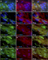
Expression of GMT-related molecular markers in Müller glial cells migrated into diabetic fibrovascular tissues. (AR) Double labeling of GS (green) and GFAP (red) (AC), TβRI (green) and GFAP (red) (DF), SNAIL (green) and GFAP (red) (GI), α-SMA (green) and GFAP (red) (JL), GFAP (green) and SM22 (red) (MO), and GS (green) and SM22 (red) (PR) in diabetic fibrovascular tissue specimens with DAPI (blue) counterstain to nuclei. Scale bar = 20 µm.
Figure 2.
Figure 2.
Autoinduction of TGFB1 expression via its noncanonical signaling pathways in Müller glial cells. (A) Müller glial cells were incubated with TGF-β1 or TGF-β2 (30 ng/mL) for 24 hours, and TGFB1 mRNA levels were analyzed. (B) Müller glial cells were pretreated with each inhibitor (U0126, ERK1/2; JSH-23, NF-κB; LY294002, PI3K; SP600125, JNK; SB203580, p38 MAPK; SB216763, GSK-3) at 10 µM for 30 minutes followed by stimulation with TGF-β1 or TGF-β2 (30 ng/mL) for 24 hours, and TGFB1 mRNA levels were analyzed. (C) SMAD2-siRNAs (siRNA-1 and siRNA-2) were transfected in Müller glial cells, and SMAD2 mRNA levels were analyzed. (D) Control siRNA-treated (Ctrl-siRNA) and SMAD2-knockdown (siRNA-1 and siRNA-2) Müller glial cells were stimulated with TGF-β1, and TGFB1 mRNA levels were analyzed. (E) Müller glial cells were incubated with TGF-β1, and protein levels of phosphorylated and total forms of NF-κB, p38, and GSK-3α/β were analyzed at various time points. (F, G) Müller glial cells were pretreated with normal IgG (300 ng/mL), anti-TβRI antibody (300 ng/mL), TβRI kinase inhibitor SB431542 (10 µg/mL), or anti-TβRII antibody (250 ng/mL) for 30 minutes followed by stimulation with TGF-β1 or TGF-β2 (30 ng/mL) for 24 hours, and TGFB1 mRNA levels were analyzed. n = 3, *P < 0.05, **P < 0.01. n.s., not significant.
Figure 3.
Figure 3.
Screening of PDR-associated profibrotic cytokines and Müller glial VEGF-A induction exclusively by TGF-β1/2. (A) Müller glial cells were applied with TGF-β1, TGF-β2, CTGF, FGF2, NGF, and PDGF-BB at the dose of 10 ng/mL for 24 hours, and VEGFA mRNA levels were analyzed. (B) Müller glial cells were incubated with TGF-β1 or TGF-β2 at different doses, and protein levels of VEGF-A were analyzed. (C, D) Müller glial cells were pretreated with normal IgG (300 ng/mL), anti-TβRI antibody (300 ng/mL), TβRI kinase inhibitor SB431542 (10 µg/mL), or anti-TβRII antibody (250 ng/mL) for 30 minutes followed by stimulation with TGF-β1 or TGF-β2 (30 ng/mL) for 24 hours, and VEGFA mRNA levels were analyzed. (E) Müller glial cells were stimulated with TGF-β1 or TGF-β2 (30 ng/mL) for 24 hours, and supernatants were collected. Human retinal microvascular endothelial cells were preincubated with anti–VEGF-A drugs bevacizumab (0.3125 mg/mL), aflibercept (0.5 mg/mL), and normal IgG (0.5 mg/mL) for 15 minutes, followed by treatment with TGF-β1/2-stimulated Müller cell culture medium for 48 hours, and processed for cell viability assay. n = 3, *P < 0.05, **P < 0.01.
Figure 4.
Figure 4.
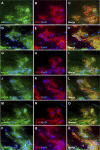
Colocalization of VEGF-A with TβRI/II in Müller glial cells migrated into diabetic fibrovascular tissues. (AO) Double labeling of GS (green) and GFAP (red) (AC), TβRI (green) and GFAP (red) (DF), VEGF-A (green) and GFAP (red) (GI), VEGF-A (green) and TβRI (red) (JL), and VEGF-A (green) and TβRII (red) (MO) in diabetic fibrovascular tissue specimens with DAPI (blue) counterstain to nuclei. Scale bar = 20 µm.
Figure 5.
Figure 5.
TGF-β1-induced VEGF-A expression via its canonical and noncanonical signaling pathways in Müller glial cells. (A) Müller glial cells were pretreated with each inhibitor (U0126, ERK1/2; JSH-23, NF-κB; LY294002, PI3K; SP600125, JNK; SB203580, p38 MAPK; SB216763, GSK-3) at 10 µM for 30 minutes followed by stimulation with TGF-β1 (30 ng/mL) for 24 hours, and VEGFA mRNA levels were analyzed. (B) Control siRNA-treated (Ctrl-siRNA) and SMAD2-knockdown (siRNA-1 and siRNA-2) Müller glial cells were stimulated with TGF-β1 (30 ng/mL) for 24 hours, and VEGFA mRNA levels were analyzed. (C, D) Müller glial cells were pretreated with each inhibitor (C) and siRNAs against SMAD2 (D) followed by stimulation with TGF-β1 (30 ng/mL) for 48 hours, and protein levels of VEGF-A were analyzed. n = 3, *P < 0.05, **P < 0.01. (E) Müller glial cells were incubated with TGF-β1 (30 ng/mL), and protein levels of phosphorylated and total forms of AKT and SMAD2 were analyzed at various time points. (F) Müller glial cells were pretreated with each inhibitor followed by stimulation with TGF-β1 (30 ng/mL) for 30 minutes, and protein levels of phosphorylated and total forms of SMAD2 were analyzed.
Figure 6.
Figure 6.
Activation of TGF-β1/2–induced signaling molecules in Müller glial cells migrated into diabetic fibrovascular tissues. (AR) Double labeling of GS (green) and GFAP (red) (AC), phosphorylated NF-κB (green) and GFAP (red) (DF), phosphorylated p38 (green) and GFAP (red) (GI), phosphorylated GSK-3 (green) and GFAP (red) (J–L), phosphorylated AKT (green) and GFAP (red) (MO) and phosphorylated SMAD2 (green) and GFAP (red) (PR) in diabetic fibrovascular tissue specimens with DAPI (blue) counterstain to nuclei. Scale bar = 20 µm.
Figure 7.
Figure 7.
Dual involvement of Müller glial autoinduction of TGF-β1/2 in diabetic fibrovascular proliferation via GMT and VEGF-A production. Müller glial autoinduction of TGF-β1/2 dually causes the pathogenic cue to facilitate and amplify diabetic fibrovascular proliferation via TGF-β1/2-driven fibrogenic GMT and VEGF-A–driven angiogenesis.

References

    1. Walshe R, Esser P, Wiedemann P, Heimann K. Proliferative retinal diseases: myofibroblasts cause chronic vitreoretinal traction. Br J Ophthalmol. 1992; 76: 550–552. - PMC - PubMed
    1. Dai Y, Wu Z, Wang F, Zhang Z, Yu M. Identification of chemokines and growth factors in proliferative diabetic retinopathy vitreous. Biomed Res Int. 2014; 2014: 486386. - PMC - PubMed
    1. Bochaton-Piallat ML, Kapetanios AD, Donati G, Redard M, Gabbiani G, Pournaras CJ. TGF-beta1, TGF-beta receptor II and ED-A fibronectin expression in myofibroblast of vitreoretinopathy. Invest Ophthalmol Vis Sci. 2000; 41: 2336–2342. - PubMed
    1. Ishida S, Shinoda K, Kawashima S, Oguchi Y, Okada Y, Ikeda E. Coexpression of VEGF receptors VEGF-R2 and neuropilin-1 in proliferative diabetic retinopathy. Invest Ophthalmol Vis Sci. 2000; 41: 1649–1656. - PubMed
    1. Hirasawa M, Noda K, Noda S, et al. .. Transcriptional factors associated with epithelial-mesenchymal transition in choroidal neovascularization. Mol Vis. 2011; 17: 1222–1230. - PMC - PubMed

Publication types

MeSH terms

Substances