. 2021 Jun;48(6):1759-1772.
doi: 10.1007/s00259-020-05133-x. Epub 2020 Dec 28.

[11C]MODAG-001-towards a PET tracer targeting α-synuclein aggregates

Affiliations

[11C]MODAG-001-towards a PET tracer targeting α-synuclein aggregates

Laura Kuebler et al. Eur J Nucl Med Mol Imaging. 2021 Jun.

Abstract

Purpose: Deposition of misfolded alpha-synuclein (αSYN) aggregates in the human brain is one of the major hallmarks of synucleinopathies. However, a target-specific tracer to detect pathological aggregates of αSYN remains lacking. Here, we report the development of a positron emission tomography (PET) tracer based on anle138b, a compound shown to have therapeutic activity in animal models of neurodegenerative diseases.

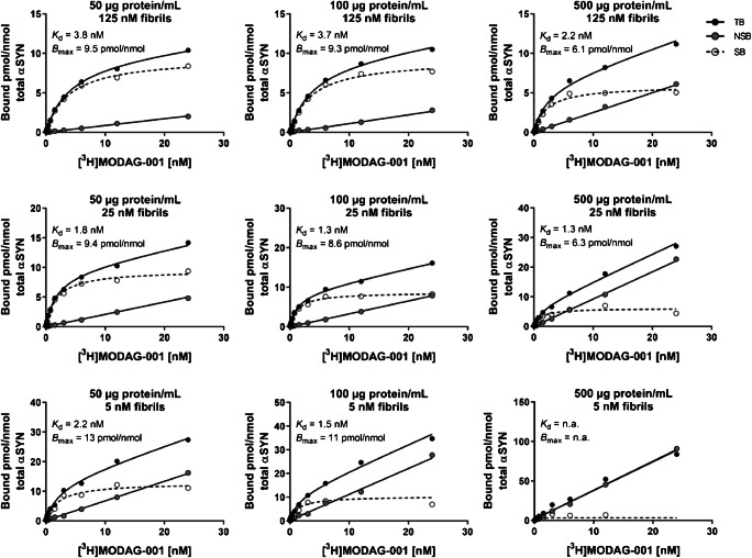
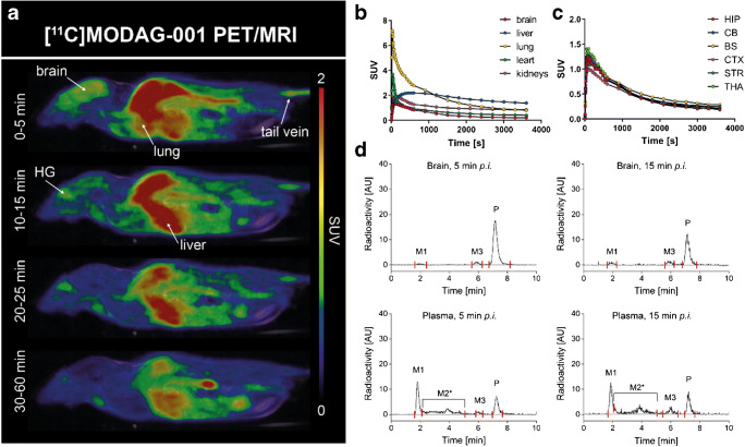
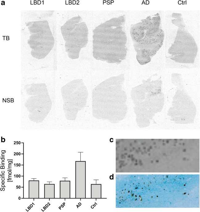
Methods: Specificity and selectivity of [3H]MODAG-001 were tested in in vitro binding assays using recombinant fibrils. After carbon-11 radiolabeling, the pharmacokinetic and metabolic profile was determined in mice. Specific binding was quantified in rats, inoculated with αSYN fibrils and using in vitro autoradiography in human brain sections of Lewy body dementia (LBD) cases provided by the Neurobiobank Munich (NBM).

Results: [3H]MODAG-001 revealed a very high affinity towards pure αSYN fibrils (Kd = 0.6 ± 0.1 nM) and only a moderate affinity to hTau46 fibrils (Kd = 19 ± 6.4 nM) as well as amyloid-β1-42 fibrils (Kd = 20 ± 10 nM). [11C]MODAG-001 showed an excellent ability to penetrate the mouse brain. Metabolic degradation was present, but the stability of the parent compound improved after selective deuteration of the precursor. (d3)-[11C]MODAG-001 binding was confirmed in fibril-inoculated rat striata using in vivo PET imaging. In vitro autoradiography showed no detectable binding to aggregated αSYN in human brain sections of LBD cases, most likely, because of the low abundance of aggregated αSYN against background protein.

Conclusion: MODAG-001 provides a promising lead structure for future compound development as it combines a high affinity and good selectivity in fibril-binding assays with suitable pharmacokinetics and biodistribution properties.

Keywords: Alpha-synuclein; PET imaging; Parkinson’s disease; Tracer development.


Conflict of interest statement

A patent has been filed.

Armin Giese, Felix Schmidt, Daniel Weckbecker, Andrei Leonov, and Sergey Ryazanov are employed by MODAG GmbH, which retains ownership of MODAG-001, and Armin Giese and Christian Griesinger are shareholders of MODAG GmbH.

Figures

Fig. 1
Fig. 1
Chemical structures and calculated logP values (clogP) of anle138b (a), anle253b (b) MODAG-001 (c), and (d3)-MODAG-001 (d) [14]
Fig. 2
Fig. 2
[3H]MODAG-001 binding experiments using recombinant human fibrils. Negative stain electron microscopy images and 4.7-fold magnification of α-synuclein (a, d), hTau46 (b, e), and amyloid-β1–42 (c, f) fibrils used in the binding experiments. Total and nonspecific binding curves of [3H]MODAG-001 to α-synuclein (g), hTau46 (h), and amyloid-β1–42 (i) fibrils. Scale bar 500 nm. TB, total binding; NSB, nonspecific binding; SB, specific binding; αSYN, α-synuclein; Aβ1–42, amyloid-β1–42
Fig. 3
Fig. 3
[3H]MODAG-001 sensitivity analysis. Determination of total binding and nonspecific binding of [3H]MODAG-001 in mouse brain homogenates spiked with recombinant α-synuclein (αSYN) fibrils. Increasing homogenate protein concentrations (left to right) were inoculated with decreasing fibril concentrations (top to bottom). At smallest concentrations of 5 nM fibrils, total binding and nonspecific curves were still separated to calculate specific binding at a homogenate concentration of 100 μg/mL, but indistinguishable at a protein concentration of 500 μg/mL. A 5-fold increase of the αSYN fibril concentration increased the total binding curve, enabling the calculation of specific binding. TB, total binding; NSB, nonspecific binding; SB, specific binding; αSYN, α-synuclein; Aβ1–42, amyloid-β1–42
Fig. 4
Fig. 4
Radiolabeling of [11C]MODAG-001. Depending on the required molar radioactivity, two different strategies were used. Direct methylation (upper reaction conditions) gave lower radiochemical yields but a high molar radioactivity (266 ± 113 MBq, 98.6 ± 24.7 GBq/μmol) while reductive methylation (lower reaction arrow) gave better yields but lower molar radioactivities (1003 ± 247 MBq, 31.3 ± 6.4 GBq/μmol)
Fig. 5
Fig. 5
Pharmacokinetic profile of [11C]MODAG-001 in the mouse. Whole-body PET/MR images of one exemplary mouse at different time points after i.v. injection (a) and corresponding time activity curves of different organs and brain regions (b, c). Images show a good brain uptake with a peak standardized uptake value of 1.4. Metabolite analysis of [11C]MODAG-001 revealed two detectable metabolites M1 and M3 present in the brain (d). PET, positron emission tomography; MRI, magnetic resonance imaging; SUV, standardized uptake value; HG, harderian glands; HIP, hippocampus; CB, cerebellum; BS, brainstem; CTX, cortex; STR, striatum; THA, thalamus; AU, arbitrary units; M1, metabolite 1; M2*, mixture of various metabolites; M3, metabolite 3; P, parent compound; p.i., post iniectio
Fig. 6
Fig. 6
Pharmacokinetic profile of (d3)-[11C]MODAG-001 in the mouse. Whole-body tracer accumulation over time is shown (a, b). (d3)-[11C]MODAG-001 rapidly entered the brain with peak standardized uptake values of 1.7 (c) followed by a fast washout. HPLC chromatograms of (d3)-[11C]MODAG-001 in plasma and brain homogenates revealed two detectable metabolites M1 and M3 present in the brain (d). HPLC, high-performance liquid chromatography, PET, positron emission tomography; MRI, magnetic resonance imaging; SUV, standardized uptake value; HG, harderian glands; HIP, hippocampus; CB, cerebellum; BS, brain stem; CTX, cortex; STR, striatum; THA, thalamus; AU, arbitrary units; M1, metabolite 1; M2*, mixture of various metabolites; M3, metabolite 3; P, parent compound; p.i., post iniectio
Fig. 7
Fig. 7
In vivo binding of (d3)-[11C]MODAG-001 in α-synuclein-inoculated rats. Coronal and transversal PET images summed up from 2.5 to 60 min of three rats (#1–3) 4 days post inoculation and one non-injected control rat (a). Images show increased tracer accumulation in the αSYN fibril-inoculated right striatum compared to the vehicle-injected contralateral striatum (a, row 1–3). Thioflavin S staining (b) confirmed the location of αSYN fibrils (white arrow) in the right striatum of fibril-inoculated rats (#1–3) (b). Time activity curves of (d3)-[11C]MODAG-001 show a rapid brain uptake with peak standardized uptake values of 2.1 ± 0.1 in the left striatum and a difference between the right (injected) and left (vehicle injected) striatum (c, d, e). The binding potential (DVR-140-60min) was 0.14 ± 0.1 for the whole striatum VOI analysis and 0.44 ± 0.21 for the 70% isocontour VOI analysis, using the contralateral side as reference region (c), while no difference was observed in the non-inoculated control rat (d). PET, positron emission tomography; αSYN, α-synuclein; rSTR, right striatum; ThS, thioflavin S; Ctrl, control; SUV, standardized uptake value, DVR-1, distribution volume ratio-1; VOI, voxel of interest
Fig. 8
Fig. 8
a [3H]MODAG-001 in vitro autoradiography (AR) on human brain slices with different pathologies: Lewy body dementia (LBD), progressive supranuclear palsy (PSP), Alzheimer’s disease (AD), healthy control (Ctrl). Images show total (TB) and nonspecific binding (NSB) of [3H]MODAG-001. b Quantification of specific tracer binding in respective brain slices in fmol/mg is shown. c Magnification of a pathological region in the AR of an AD case counterstained for Aβ plaques (d)

References

    1. Spillantini MG, Schmidt ML, Lee VM, Trojanowski JQ, Jakes R, Goedert M. Alpha-synuclein in Lewy bodies. Nature. 1997;388(6645):839–840. doi: 10.1038/42166.. - DOI - PubMed
    1. Fanciulli A, Wenning GK. Multiple-system atrophy. N Engl J Med. 2015;372(14):1375–1376. doi: 10.1056/NEJMc1501657. - DOI - PubMed
    1. Graham JG, Oppenheimer DR. Orthostatic hypotension and nicotine sensitivity in a case of multiple system atrophy. J Neurol Neurosurg Psychiatry. 1969;32(1):28–34. doi: 10.1136/jnnp.32.1.28. - DOI - PMC - PubMed
    1. Gibb WR, Esiri MM, Lees AJ. Clinical and pathological features of diffuse cortical Lewy body disease (Lewy body dementia) Brain. 1987;110(Pt 5):1131–1153. doi: 10.1093/brain/110.5.1131. - DOI - PubMed
    1. Gomez-Tortosa E, Newell K, Irizarry MC, Sanders JL. Hyman BT. alpha-Synuclein immunoreactivity in dementia with Lewy bodies: morphological staging and comparison with ubiquitin immunostaining. Acta Neuropathol. 2000;99(4):352–357. doi: 10.1007/s004010051135.. - DOI - PubMed

Publication types