Review
. 2021 Jan 1;147(1):54e-65e.
doi: 10.1097/PRS.0000000000007451.

Craniofacial and Long Bone Development in the Context of Distraction Osteogenesis

Affiliations
Review

Craniofacial and Long Bone Development in the Context of Distraction Osteogenesis

Harsh N Shah et al. Plast Reconstr Surg. .

Abstract

Background: Bone retains regenerative potential into adulthood, and surgeons harness this plasticity during distraction osteogenesis. The underlying biology governing bone development, repair, and regeneration is divergent between the craniofacial and appendicular skeleton. Each type of bone formation is characterized by unique molecular signaling and cellular behavior. Recent discoveries have elucidated the cellular and genetic processes underlying skeletal development and regeneration, providing an opportunity to couple biological and clinical knowledge to improve patient care.

Methods: A comprehensive literature review of basic and clinical literature regarding craniofacial and long bone development, regeneration, and distraction osteogenesis was performed.

Results: The current understanding in craniofacial and long bone development and regeneration is discussed, and clinical considerations for the respective distraction osteogenesis procedures are presented.

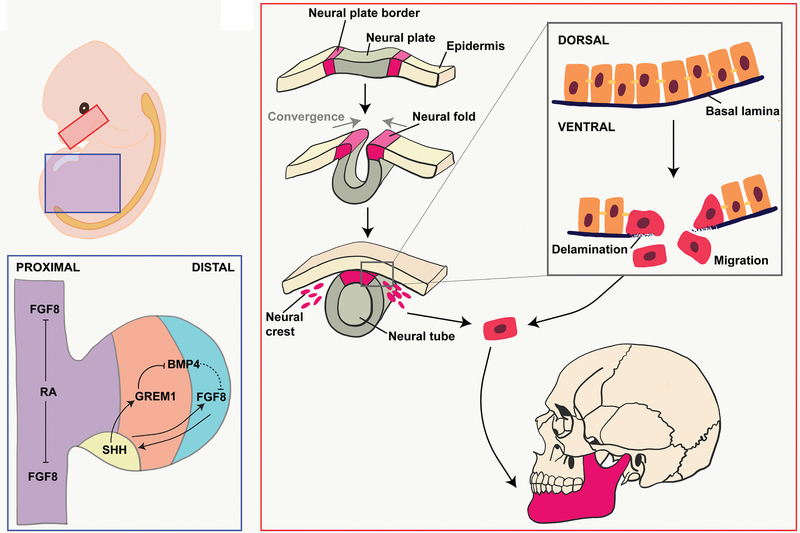
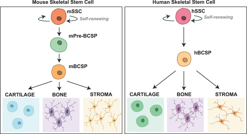
Conclusions: Distraction osteogenesis is a powerful tool to regenerate bone and thus address a number of craniofacial and appendicular skeletal deficiencies. The molecular mechanisms underlying bone regeneration, however, remain elusive. Recent work has determined that embryologic morphogen gradients constitute important signals during regeneration. In addition, striking discoveries have illuminated the cellular processes underlying mandibular regeneration during distraction osteogenesis, showing that skeletal stem cells reactivate embryologic neural crest transcriptomic processes to carry out bone formation during regeneration. Furthermore, innovative adjuvant therapies to complement distraction osteogenesis use biological processes active in embryogenesis and regeneration. Additional research is needed to further characterize the underlying cellular mechanisms responsible for improved bone formation through adjuvant therapies and the role skeletal stem cells play during regeneration.


Figures

Figure 1.
Figure 1.. Embryonic View of Bone Development.
(A) Developing mandible (red box) and lower limb (blue box). (B) Endothelial-to-mesenchymal transition of cranial neural crest cells. (C) Signaling throughout the limb bud trunk (purple), zone of polarizing activity (yellow), progress zone (orange), and apical endodermal ridge (blue).
Figure 2.
Figure 2.. Tyrosine Kinase Pathway.
The single pass, type I receptor tyrosine kinase resides in the plasma membrane. The receptor tyrosine kinase is activated through the binding of a ligand leading to a ligand-induced dimerization with the cytoplasmic tyrosine kinase domain. The dimerization results in autophosphorylation of the tyrosine residues inducing conformational changes which stabilize the active site of the kinase. The phosphotyrosine residues act as recruitment sites for downstream signaling proteins.
Figure 3.
Figure 3.. Hoxa2/Runx2 Pathway.
(A) Mouse embryo with developing pharyngeal arches (PA). PA1 (red) gives rise to the muscles of mastication and mandible. PA2 (orange) gives rise to the muscles of facial expression and hyoid bone. PA3 (yellow) gives rise to the greater horn and lower body of the hyoid bone. PA4 (green) gives rise to the thyroid and cricoid cartilage. (B) In the absence of Hoxa2, Runx2 activation will occur leading to bone formation. In the presence of Hoxa2, Runx2 will be suppressed limiting bone formation. (C) The expression of Hoxa2 increases in the caudal direction of the pharyngeal arches with PA1 not having expression of Hoxa2, while PA4 possesses a high level of Hoxa2 expression. Analogously, the expression of Runx2 decreases in the caudal direction of the pharyngeal arches with PA1 having the greatest expression of Runx2, while PA4 possesses a low level of Runx2 expression.
Figure 4.
Figure 4.. Skeletal Stem Cell Hierarchy.
(A) The skeletal stem cell hierarchy in mice beginning with a self-renewing mouse Skeletal Stem Cell (mSSC) differentiating to lineage-restricted bone, cartilage, and stromal cells through a Bone, Cartilage, Stromal Progenitor (BCSP) cell. (B) The skeletal stem cell hierarchy in humans beginning with a self-renewing human Skeletal Stem Cell (hSSC) differentiating to lineage-restricted bone, cartilage, and stromal cells through a Bone, Cartilage, Stromal Progenitor (BCSP) cell.
Figure 5.
Figure 5.. Focal Adhesion Kinase Signaling Pathway.
Cytoplasmic tyrosine kinase FAK becomes activated after interacting with transmembrane integrin proteins allowing FAK to form a complex with Src family kinase. The complex initiates downstream signaling pathways through the phosphorylation of other proteins such as ERK/MAPK.

References

    1. Orioli IM, Castilla EE, Barbosa-Neto JG. The birth prevalence rates for the skeletal dysplasias. J Med Genet. 1986;23(4):328–332. - PMC - PubMed
    1. Krakow D Skeletal dysplasias. Clin Perinatol. 2015;42(2):301–319, viii. - PMC - PubMed
    1. Cauley JA. Public health impact of osteoporosis. J Gerontol A Biol Sci Med Sci. 2013;68(10):1243–1251. - PMC - PubMed
    1. Kanis JA, Oden A, Johansson H, Borgstrom F, Strom O, McCloskey E. FRAX and its applications to clinical practice. Bone. 2009;44(5):734–743. - PubMed
    1. Unnanuntana A, Gladnick BP, Donnelly E, Lane JM. The assessment of fracture risk. J Bone Joint Surg Am. 2010;92(3):743–753. - PMC - PubMed