. 2020 Dec 21;25(24):6045.
doi: 10.3390/molecules25246045.

Therapeutic Potential of Rhododendron arboreum Polysaccharides in an Animal Model of Lipopolysaccharide-Inflicted Oxidative Stress and Systemic Inflammation

Affiliations

Therapeutic Potential of Rhododendron arboreum Polysaccharides in an Animal Model of Lipopolysaccharide-Inflicted Oxidative Stress and Systemic Inflammation

Ajaz Ahmad et al. Molecules. .

Abstract

Systemic inflammation results in physiological changes, largely mediated by inflammatory cytokines. The present investigation was performed to determine the effect of Rhododendron arboreum (RAP) on inflammatory parameters in the animal model. The RAP (100 and 200 mg/kg) were pre-treated for animals, given orally for one week, followed by lipopolysaccharide (LPS) injection. Body temperature, burrowing, and open field behavioral changes were assessed. Biochemical parameters (AST, ALT, LDH, BIL, CK, Cr, BUN, and albumin) were done in the plasma after 6 h of LPS challenge. Oxidative stress markers SOD, CAT, and MDA were measured in different organs. Levels of inflammatory markers like tumor necrosis factor-alpha (TNF-α), interleukin-1-beta (IL-1β) and, interleukin-6 (IL-6) as well as VEGF, a specific sepsis marker in plasma, were quantified. The plasma enzymes, antioxidant markers and plasma pro-inflammatory cytokines were significantly restored (p < 0.5) by RAP treatment, thus preventing the multi-organ and tissue damage in LPS induced rats. The protective effect of RAP may be due to its potent antioxidant potential. Thus, RAP can prevent LPS induced oxidative stress, as well as inflammatory and multi-organ damage as reported in histopathological studies in rats when administered to the LPS treated animals. These findings indicate that RAP can benefit in the management of systemic inflammation from LPS and may have implications for a new treatment or preventive therapeutic strategies with an inflammatory component.

Keywords: Rhododendron arboreum polysaccharides; animal model; lipopolysaccharide; multi-biomarker approach; oxidative stress.


Conflict of interest statement

The authors declare no conflict of interest.

Figures

Figure 1
Figure 1
Determination of SOD in (A) plasma, (B) Liver and (C) Brain. Results are mean SEM of seven animals per group. ‘*’ denotes significant differences compared with the normal control group (p < 0.05); ‘#’ denotes significant differences compared with the LPS group (p < 0.05).
Figure 2
Figure 2
Determination of CAT in (A) plasma, (B) Liver and (C) Brain. Results are mean SEM of seven animals per group. ‘*’ denotes significant differences compared with the normal control group (p < 0.05); ‘#’ denotes significant differences compared with the LPS group (p < 0.05).
Figure 3
Figure 3
Determination of MDA in (A) plasma, (B) Liver and (C) Brain. Results are mean SEM of seven animals per group. ‘*’ denotes significant differences compared with the normal control group (p < 0.05); ‘#’ denotes significant differences compared with the LPS group (p < 0.05).
Figure 4
Figure 4
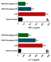
Effect of RAP (100 and 200 mg/kg) on pro-inflammatory cytokine levels in the plasma of LPS-induced systemic inflammation. (A) Tumor necrosis factor-a (TNF-α), and (B) Interleukin-1β (IL-1β). Results are mean SEM of seven animals per group. ‘*’ denotes significant differences compared with the normal control group (p < 0.05); ‘#’ denotes significant differences compared with the LPS group (p < 0.05).
Figure 5
Figure 5
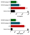
Effect of RAP (100 and 200 mg/kg) on pro-inflammatory cytokine levels in the plasma of LPS-induced systemic inflammation (A) Interleukin-6 (IL-6), and (B) Vascular endothelial growth factor (VEGF); results are mean SEM of seven animals per group. ‘*’ denotes significant differences compared with the normal control group (p < 0.05); ‘#’ denotes significant differences compared with the LPS group (p < 0.05).
Figure 6
Figure 6
Effect of RAP (100 and 200 mg/kg) on histological alterations in the liver of LPS-induced systemic inflammation. Normal control rats showing intact hepatocytes and normal histological structure of central vein. LPS treated rats showing multiple focal necrosis, severe damage of hepatic architecture, and damage in hepatocytes. Rats treated with RAP 100 mg/kg + LPS and RAP 200 mg/kg + LPS showing absence of histopatholgical alterations.
Figure 7
Figure 7
Effect of RAP (100 and 200 mg/kg) on histological alterations in the brain of LPS-induced systemic inflammation. There is a severe loss of intact cells in the cortical region of the brain. LPS group shows more vacuolated and edematous tissue in comparison to the normal control group. RAP administration has shown a decrease in neuronal loss. The normal control group has shown the normal morphology of the cortical region.

References

    1. Medzhitov R. Inflammation 2010: New Adventures of an Old Flame. Cell. 2010;140:771–776. doi: 10.1016/j.cell.2010.03.006. - DOI - PubMed
    1. Angus D.C., Linde-Zwirble W.T., Lidicker J., Clermont G., Carcillo J., Pinsky M.R. Epidemiology of severe sepsis in the United States: Analysis of incidence, outcome, and associated costs of care. Crit. Care Med. 2001;29:1303–1310. doi: 10.1097/00003246-200107000-00002. - DOI - PubMed
    1. Stearns-Kurosawa D., Osuchowski M.F., Valentine C., Kurosawa S., Remick D.G. The Pathogenesis of Sepsis. Annu. Rev. Pathol. Mech. Dis. 2011;6:19–48. doi: 10.1146/annurev-pathol-011110-130327. - DOI - PMC - PubMed
    1. Marx G., Vangerow B., Burczyk C., Gratz K., Maassen N., Meyer M.C., Leuwer M., Kuse E., Rueckoldt H. Evaluation of noninvasive determinants for capillary leakage syndrome in septic shock patients. Intensiv. Care Med. 2000;26:1252–1258. doi: 10.1007/s001340000601. - DOI - PubMed
    1. Van Der Flier M., Van Leeuwen H.J., Van Kessel K.P., Kimpen J.L., Hoepelman A.I., Geelen S.P. Plasma vascular endothelial growth factor in severe sepsis. Shock. 2005;23:35–38. doi: 10.1097/01.shk.0000150728.91155.41. - DOI - PubMed