Review
. 2020 Dec 21;12(12):3061.
doi: 10.3390/polym12123061.

Biopolymer Coatings for Biomedical Applications

Affiliations
Review

Biopolymer Coatings for Biomedical Applications

A Joseph Nathanael et al. Polymers (Basel). .

Abstract

Biopolymer coatings exhibit outstanding potential in various biomedical applications, due to their flexible functionalization. In this review, we have discussed the latest developments in biopolymer coatings on various substrates and nanoparticles for improved tissue engineering and drug delivery applications, and summarized the latest research advancements. Polymer coatings are used to modify surface properties to satisfy certain requirements or include additional functionalities for different biomedical applications. Additionally, polymer coatings with different inorganic ions may facilitate different functionalities, such as cell proliferation, tissue growth, repair, and delivery of biomolecules, such as growth factors, active molecules, antimicrobial agents, and drugs. This review primarily focuses on specific polymers for coating applications and different polymer coatings for increased functionalization. We aim to provide broad overview of latest developments in the various kind of biopolymer coatings for biomedical applications, in order to highlight the most important results in the literatures, and to offer a potential outline for impending progress and perspective. Some key polymer coatings were discussed in detail. Further, the use of polymer coatings on nanomaterials for biomedical applications has also been discussed, and the latest research results have been reported.

Keywords: bioactivity; biomedical applications; biopolymers; coatings; nanoparticles; tissue engineering.


Conflict of interest statement

The authors declare no conflict of interest.

Figures
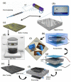
Figure 1
Figure 1
(a) electrospinning process, (b) photograph of the large-scale prepared mat of highly aligned PVDF NFs arrays; enlarged view exhibits the structure from respective section, (c) structure of oxidant-contained yellowish PVDF NFs mat before PANI coating, (d) VPP process, (e) structure of deepbluish PVDF NFs mat after PANI coating, (f) electrode assembling, (g) lamination process, (h) PDMS encapsulation of POESS design, (i) photographs of POESS with demonstration of flexibility. Schematic illustration of the piezo-organic-e-skin sensor design architecture. (Reprinted with permission from [45] Copyright (2020), American Chemical Society.).
Figure 2
Figure 2
Viability of (a) HeLa cells and (b) MC3T3 cells on PVDF and PVDF:CP electrospun fibers (control: PVDF). ** and **** signifies p < 0.01 (1d) and p < 0.0001 (1d), respectively, for both HeLa and MC3T3 culture (1d); #### signifies p < 0.0001 (3d). CP, conducting polymers; PVDF, poly(vinylidene fluoride) (Reprinted with permission from [47] Copyright (2020), John Wiley and Sons.)
Figure 3
Figure 3
Multiscale mechanical analysis of polymethyl methacrylate layers grafted on Ti substrates. (Reprinted with permission from [14] Copyright (2017), Elsevier).
Figure 4
Figure 4
(a) Schematic illustration of polymethyl methacrylate/chitosan-silver (PMMA/AgNPs-CS) coating and its antibacterial activity; (b) optical density of suspension of Escherichia coli and Staphylococcus aureus. (Reprinted with permission from [56] Copyright (2019), Elsevier).
Figure 5
Figure 5
Scanning electron microscopy (A,B) and atomic force microscopy (C,D) images of poly(styryl bisphosphonate) (poly(StBP))-6, and poly(StBP)-40 (D) films, respectively, where “6” and “40” represent the thickness of the Mayer rod (6 and 40 µm) used to spread the polymer solution. (Reprinted with permission from [62] Copyright (2020), Elsevier).
Figure 6
Figure 6
Schematic illustration of polydimethylsiloxane (PDMS) coating using engineered anchor peptides fused to the cell-adhesive peptide sequence (glycine-arginine-glycine-aspartateserine, GRGDS). (Reprinted with permission from [63] Copyright (2019), American Chemical Society).
Figure 7
Figure 7
Schematic illustration of polydimethylsiloxane (PDMS) surface modification using hyaluronic acid and polydopamine (HA/PDA) composite coatings. (Reprinted with permission from [67]. Copyright (2017), American Chemical Society).
Figure 8
Figure 8
Schematic illustration of the cell trapping mechanism in polydimethylsiloxane (PDMS) microwells and the corresponding micrographs in (a) 200 µm and (b) 35 µm square microwell arrays. (c) Single-cell trapping demonstrated using a combination of bright field microscopy and fluorescence imaging. (Reprinted with permission from [68] Copyright (2018), American Institute of Physics).
Figure 9
Figure 9
Different approaches of chitosan coatings for nanoparticles. (Reprinted with permission from [90] Copyright (2020), Elsevier).
Figure 10
Figure 10
Schematic illustration of iron oxide nanorods coated with linear bisphosphonate−poly(ethylene glycol) (OPT), polyacrylic sodium salt (PAA), and polymethacrylate backbone/PEG side chain comb polymer (PCP). (Reprinted with permission from [97]. Copyright (2018), American Chemical Society).
Figure 11
Figure 11
Schematic illustration of imatinib mesylate (IM)-loaded PSS/PEI-AuNPs delivering IM to the layers of skin in melanoma treatment. (Reprinted with permission from [102]. Copyright (2015), American Chemical Society).

References

    1. Makhlouf A.S.H., Perez A., Guerrero E. Advances in Smart Coatings and Thin Films for Future Industrial and Biomedical Engineering Applications. Elsevier Inc.; Amsterdam, The Netherland: 2019. Recent trends in smart polymeric coatings in biomedicine and drug delivery applications; pp. 359–381. - DOI
    1. Augello C., Liu H. Surface Modification of Magnesium and its Alloys for Biomedical Applications. Volume 2. Elsevier Ltd.; Amsterdam, The Netherlands: 2015. Surface modification of magnesium by functional polymer coatings for neural applications; pp. 335–353. - DOI
    1. Landry M.J., Rollet F.G., Kennedy T.E., Barrett C.J. Layers and Multilayers of Self-Assembled Polymers: Tunable Engineered Extracellular Matrix Coatings for Neural Cell Growth. Langmuir. 2018;34:8709–8730. doi: 10.1021/acs.langmuir.7b04108. - DOI - PubMed
    1. Park S., Han U., Choi D., Hong J. Layer-by-layer assembled polymeric thin films as prospective drug delivery carriers: Design and applications. Biomater. Res. 2018;22:29. doi: 10.1186/s40824-018-0139-5. - DOI - PMC - PubMed
    1. Leontidis E. Langmuir-Blodgett Films: Sensor and Biomedical Applications and Comparisons with the Layer-by-Layer Method. In: Gursoy M., Karaman M., editors. Surface Treatments for Biological, Chemical and Physical Applications. Wiley-VCH Verlag; Weinheim, Germany: 2016. pp. 181–208. - DOI

LinkOut - more resources