

Exploring the Anti-Cancer Mechanism of Novel 3,4'-Substituted Diaryl Guanidinium Derivatives
- PMID: 33371382
- PMCID: PMC7767381
- DOI: 10.3390/ph13120485
Exploring the Anti-Cancer Mechanism of Novel 3,4'-Substituted Diaryl Guanidinium Derivatives
Abstract
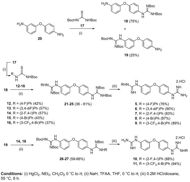
We previously identified a guanidinium-based lead compound that inhibited BRAF through a hypothetic type-III allosteric mechanism. Considering the pharmacophore identified in this lead compound (i.e., "lipophilic group", "di-substituted guanidine", "phenylguanidine polar end"), several modifications were investigated to improve its cytotoxicity in different cancer cell lines. Thus, several lipophilic groups were explored, the di-substituted guanidine was replaced by a secondary amine and the phenyl ring in the polar end was substituted by a pyridine. In a structure-based design approach, four representative derivatives were docked into an in-house model of an active triphosphate-containing BRAF protein, and the interactions established were analysed. Based on these computational studies, a variety of derivatives was synthesized, and their predicted drug-like properties calculated. Next, the effect on cell viability of these compounds was assessed in cell line models of promyelocytic leukaemia and breast, cervical and colorectal carcinomas. The potential of a selection of these compounds as apoptotic agents was assessed by screening in the promyelocytic leukaemia cell line HL-60. The toxicity against non-tumorigenic epithelial MCF10A cells was also investigated. These studies allowed for several structure-activity relationships to be derived. Investigations on the mechanism of action of representative compounds suggest a divergent effect on inhibition of the MAPK/ERK signalling pathway.
Keywords: 3,4′-bis-guanidino; 3-amino-4′-guanidino; BRAF; HL-60; apoptosis; cancer cell viability; diphenyl ether; intramolecular hydrogen bond; phenyl pyridyl ether.
Conflict of interest statement
The authors declare no conflict of interest.
Figures


Similar articles
-
Effect of isouronium/guanidinium substitution on the efficacy of a series of novel anti-cancer agents.Medchemcomm. 2018 Mar 27;9(4):735-743. doi: 10.1039/c8md00089a. eCollection 2018 Apr 1. Medchemcomm. 2018. PMID: 30108964 Free PMC article.
-
Guanidinium-based derivatives: searching for new kinase inhibitors.Eur J Med Chem. 2014 Jun 23;81:427-41. doi: 10.1016/j.ejmech.2014.05.025. Epub 2014 May 9. Eur J Med Chem. 2014. PMID: 24858546
-
Design, synthesis, in vitro antiproliferative activity and apoptosis-inducing studies of 1-(3',4',5'-trimethoxyphenyl)-3-(2'-alkoxycarbonylindolyl)-2-propen-1-one derivatives obtained by a molecular hybridisation approach.J Enzyme Inhib Med Chem. 2018 Dec;33(1):1225-1238. doi: 10.1080/14756366.2018.1493473. J Enzyme Inhib Med Chem. 2018. PMID: 30141353 Free PMC article.
-
Synthesis and Evaluation of 2-Naphthaleno trans-Stilbenes and Cyanostilbenes as Anticancer Agents.Anticancer Agents Med Chem. 2018;18(4):556-564. doi: 10.2174/1871521409666170412115703. Anticancer Agents Med Chem. 2018. PMID: 28403783 Free PMC article.
-
Cephalostatin analogues--synthesis and biological activity.Fortschr Chem Org Naturst. 2004;87:1-80. doi: 10.1007/978-3-7091-0581-8_1. Fortschr Chem Org Naturst. 2004. PMID: 15079895 Review.
Cited by
-
Special Issue "Anticancer Drugs 2021".Pharmaceuticals (Basel). 2022 Apr 14;15(4):479. doi: 10.3390/ph15040479. Pharmaceuticals (Basel). 2022. PMID: 35455476 Free PMC article.
-
Antibacterial activity of isopropoxy benzene guanidine against Riemerella anatipestifer.Front Pharmacol. 2024 Feb 2;15:1347250. doi: 10.3389/fphar.2024.1347250. eCollection 2024. Front Pharmacol. 2024. PMID: 38370472 Free PMC article.
References
Grants and funding
LinkOut - more resources
Full Text Sources
Research Materials
Miscellaneous
