. 2020 Dec 21;12(12):3869.
doi: 10.3390/cancers12123869.

Breast Cancer Organoids Model Patient-Specific Response to Drug Treatment

Affiliations

Breast Cancer Organoids Model Patient-Specific Response to Drug Treatment

Elena Campaner et al. Cancers (Basel). .

Abstract

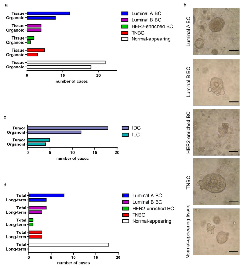
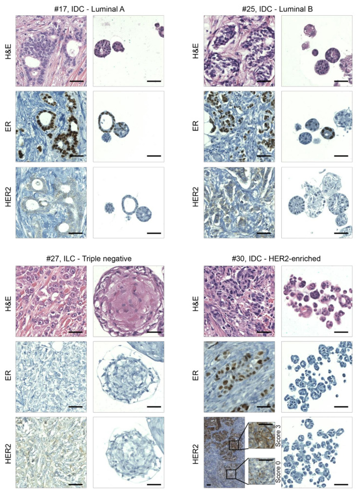
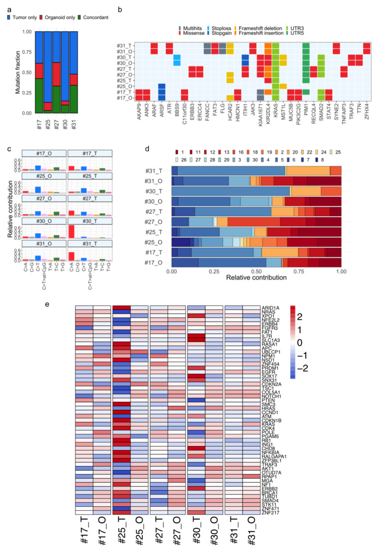
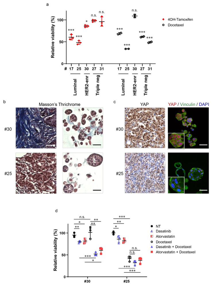
Tumor organoids are tridimensional cell culture systems that are generated in vitro from surgically resected patients' tumors. They can be propagated in culture maintaining several features of the tumor of origin, including cellular and genetic heterogeneity, thus representing a promising tool for precision cancer medicine. Here, we established patient-derived tumor organoids (PDOs) from different breast cancer subtypes (luminal A, luminal B, human epidermal growth factor receptor 2 (HER2)-enriched, and triple negative). The established model systems showed histological and genomic concordance with parental tumors. However, in PDOs, the ratio of diverse cell populations was frequently different from that originally observed in parental tumors. We showed that tumor organoids represent a valuable system to test the efficacy of standard therapeutic treatments and to identify drug resistant populations within tumors. We also report that inhibitors of mechanosignaling and of Yes-associated protein 1 (YAP) activation can restore chemosensitivity in drug resistant tumor organoids.

Keywords: YAP; breast cancer; dasatinib; drug testing; heterogeneity; mechanotransduction; patient-derived tumor organoids; statin.


Conflict of interest statement

The authors declare no conflict of interest.

Figures

Figure 1
Figure 1
Generating a collection of organoids from primary breast cancers (BCs). (a) Histogram representing the stratification of the collected BC, and the derived organoids, based on the molecular subtype. The number of collected normal-appearing adjacent-to-tumor (NAT) specimens and derived organoids is also shown. (b) Representative images of organoids derived from BC of different molecular subtypes and from NAT tissue. Scale bar, 100 μm. (c) Histogram representing the stratification of the collected BC, and the derived organoids, referring to the histological subtype; IDC, invasive ductal carcinoma; ILC, invasive lobular carcinoma. (d) Histogram depicting the number of PDOs maintained in culture for longer time (Long-term) compared with the total number of established cultures, arranged into the molecular subtype. The number of long-term NAT-derived organoids is also shown. Long-term organoids are defined as organoids maintained for more than 4 weeks in culture (passages ≥ 4). TNBC, triple negative BC; HER2, human epidermal growth factor receptor 2.
Figure 2
Figure 2
Organoids recapitulate the histological features of primary BCs. Representative images of hematoxylin/eosin staining (H&E) and immunohistochemical analyses on sections of BCs and derived organoids. From left to right and from the top to the bottom are examples of luminal A, luminal B, triple negative, and HER2-enriched BCs, respectively; IDC, invasive ductal carcinoma; ILC, invasive lobular carcinoma; ER, estrogen receptor; HER2, human epidermal growth factor receptor 2. Magnifications of HER2 staining in different areas of cancer tissue #30 are also shown. Scale bar, 100 μm.
Figure 3
Figure 3
Comparative analysis of mutational signature and cancer genes in BC tumor–organoid pairs. (a) Stacked bar chart showing the distribution of somatic mutations specific to tumor tissues (blue) and organoids (red), across five tumor–organoid pairs. The proportion of common mutations between tumor and matched organoid model is indicated in green. (b) Overview of somatic nonsynonymous mutations, also affecting cancer genes, shared in tumor and paired organoids. (c) Bar graphs representing the relative contribution of the indicated mutation types to the mutational signature extracted by non-negative matrix factorization (NMF) analysis from whole-exome sequencing (WES) data, for each tumor and organoid sample. (d) The COSMIC single-base substitution (SBS) signatures (1–30) are conserved among matching tumor–organoid pairs, most of which are annotated as associated to BC (i.e., 1, 2, 3, 5, 13). (e) Heat map comparing copy number variations across tumor–organoid pairs in a log2 scale, in BC relevant genes. Red colors indicate gains, and blue colors indicate losses. Samples are labeled as tumor (T) and organoid (O). UTR, untranslated region.
Figure 4
Figure 4
Tumor organoids represent a powerful model to evaluate novel potential therapeutic strategies. (a) Dot plot showing relative viability of BC organoids treated with either 10 μM 4-Hydroxytamoxifen (4OH-Tamoxifen) or 1 nM Docetaxel for 7 days. Data are normalized to control treatment (solvent), set to 100%. Ethanol was used as control for 4-Hydroxytamoxifen, while dimethyl sulfoxide (DMSO) was used for Docetaxel. Data shown are the means ± s.d. of n = 3 (4OH-Tamoxifen) and n = 2 (Docetaxel) independent experiments; * p < 0.05, *** p < 0.001; n.s., not significant; t-test, using the Holm–Sidak method. (b) Representative images of Masson’s trichrome staining on sections of BC (left) and derived organoids (right) of the indicated cases. Scale bar, 50 μm. (c) Representative images of Yes-associated protein 1 (YAP) immunohistochemistry on BC sections (left) and of YAP (red) and Vinculin (green) immunofluorescence on sections of the derived organoids of the same cases (right). Nuclei were counterstained with either hematoxylin (left) or 4′,6-diamidino-2-phenylindole (DAPI) (right). Scale bar, 50 μm. Magnifications of YAP/Vinculin staining are also shown. (d) Dot plot showing relative viability of BC organoids treated with 100 nM Dasatinib, 100 nM Atorvastatin, 1 nM Docetaxel, their combinations, or solvent (NT, DMSO) for 7 days. Data shown are the means ± s.d. of n = 3 independent experiments, * p < 0.05, ** p < 0.01, *** p < 0.001; n.s., not significant; t-test, using the Holm–Sidak method.

References

    1. Dagogo-Jack I., Shaw A.T. Tumour heterogeneity and resistance to cancer therapies. Nat. Rev. Clin. Oncol. 2018;15:81–94. doi: 10.1038/nrclinonc.2017.166. - DOI - PubMed
    1. Zardavas D., Irrthum A., Swanton C., Piccart M. Clinical management of breast cancer heterogeneity. Nat. Rev. Clin. Oncol. 2015;12:381–394. doi: 10.1038/nrclinonc.2015.73. - DOI - PubMed
    1. Leight J.L., Drain A.P., Weaver V.M. Extracellular Matrix Remodeling and Stiffening Modulate Tumor Phenotype and Treatment Response. Annu. Rev. Cancer Biol. 2017;1:313–334. doi: 10.1146/annurev-cancerbio-050216-034431. - DOI
    1. velaei K., Samadi N., Barazvan B., Soleimani Rad J. Tumor microenvironment-mediated chemoresistance in breast cancer. The Breast. 2016;30:92–100. doi: 10.1016/j.breast.2016.09.002. - DOI - PubMed
    1. Hirata E., Sahai E. Tumor Microenvironment and Differential Responses to Therapy. Cold Spring Harb. Perspect. Med. 2017;7:a026781. doi: 10.1101/cshperspect.a026781. - DOI - PMC - PubMed