Review
. 2020 Dec 21;21(24):9769.
doi: 10.3390/ijms21249769.

Melanogenesis Connection with Innate Immunity and Toll-Like Receptors

Affiliations
Review

Melanogenesis Connection with Innate Immunity and Toll-Like Receptors

Saaya Koike et al. Int J Mol Sci. .

Abstract

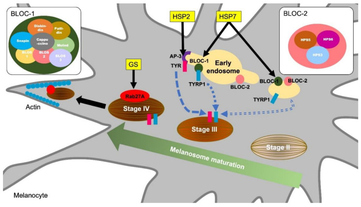
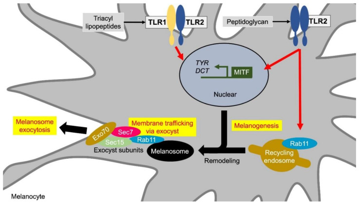
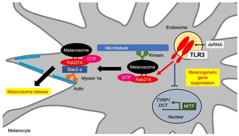
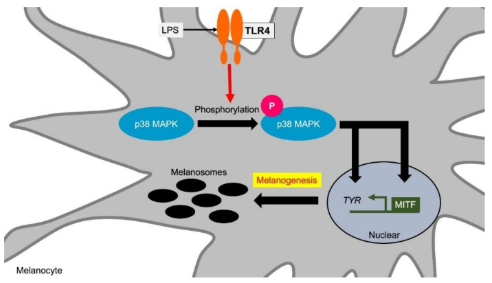
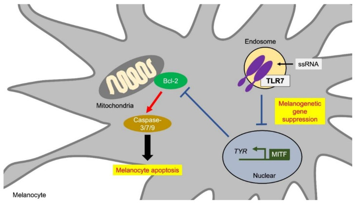
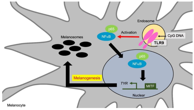
The epidermis is located in the outermost layer of the living body and is the place where external stimuli such as ultraviolet rays and microorganisms first come into contact. Melanocytes and melanin play a wide range of roles such as adsorption of metals, thermoregulation, and protection from foreign enemies by camouflage. Pigmentary disorders are observed in diseases associated with immunodeficiency such as Griscelli syndrome, indicating molecular sharing between immune systems and the machineries of pigment formation. Melanocytes express functional toll-like receptors (TLRs), and innate immune stimulation via TLRs affects melanin synthesis and melanosome transport to modulate skin pigmentation. TLR2 enhances melanogenetic gene expression to augment melanogenesis. In contrast, TLR3 increases melanosome transport to transfer to keratinocytes through Rab27A, the responsible molecule of Griscelli syndrome. TLR4 and TLR9 enhance tyrosinase expression and melanogenesis through p38 MAPK (mitogen-activated protein kinase) and NFκB signaling pathway, respectively. TLR7 suppresses microphthalmia-associated transcription factor (MITF), and MITF reduction leads to melanocyte apoptosis. Accumulating knowledge of the TLRs function of melanocytes has enlightened the link between melanogenesis and innate immune system.

Keywords: Griscelli syndrome; Hermansky–Pudlak syndrome; RAB; innate immunity; melanogenesis; melanosome; microphthalmia-associated transcription factor; toll-like receptor; tyrosinase.


Conflict of interest statement

The authors declare no conflict of interest.

Figures

Figure 1
Figure 1
Molecular mechanisms of melanosome formation and maturation related to the hypopigmentary genetic diseases. Griscelli syndrome (GS) is an autosomal recessive disorder characterized by immunodeficiency and partial bleaching due to abnormal melanosome distribution. One of the causes of GS is a mutation in the Rab27A gene. Rab27A transports melanosome to the cell periphery, and Rab27A gene mutations result in a melanosome transport disturbance in melanocytes. Hermansky–Pudlak syndromes (HPS) are also rare autosomal recessive disorders and are associated with skin depigmentation and immune impairment. HPS2 patients have a deficiency in the β3A subunit of AP-3. AP-3 classifies the melanin synthesis-related enzyme tyrosinase (TYR) from endosomes to melanosomes. Patients with HSP7 do not express the dysbindin protein, one of the components of BLOC-1 (biogenesis of lysosome-related organelles complex 1). BLOC-1 regulates the transport and biosynthesis of lysosomal organelles. BLOC-1 interacts with AP-3 to promote the transport of tyrosinase-related protein 1 (TYRP1). BLOC-1 also interacts with BLOC-2 and promotes TYRP1 transport by a mechanism different from that of AP-3.
Figure 2
Figure 2
TLR1/2 and TLR2 induce melanin formation and melanosome exocytosis. Stimuli for the TLR1/2 heterodimer and the TLR2 homodimer promote melanin synthesis by inducing the transcription of melanin synthesis-related genes such as MITF, TYR, and DCT. In addition, TLR2 induces extracellular release of melanosomes by increasing the expression of Rab11. Rab11 initiates exocytosis through the remodeling of mature melanosome membranes. Rab11 also constitutes a subunit with tethering factors such as Sec8, Sec15, and Exo70, and anchors melanosomes to the cell membrane. Abbreviations: TLR: toll-like receptor; MITF: microphthalmia-associated transcription factor; TYR: tyrosinase; DCT: dopachrome tautomerase.
Figure 3
Figure 3
TLR3 promotes melanosome transfer. TLR3 stimulation reduces de novo melanin synthesis by suppressing the expression of MITF, TYRP1, and DCT. On the other hand, TLR3 stimulation promotes extracellular release of melanosomes through Rab27A induction. Rab27A on melanosomes binds to the actin-dependent motor protein myosin Va via Slac2-a and promotes actin transport toward the cell periphery near the cell membrane by forming a tripartite complex. Abbreviations: TLR: toll-like receptor; MITF: microphthalmia-associated transcription factor; TYRP1: tyrosinase-related protein 1; DCT: dopachrome tautomerase.
Figure 4
Figure 4
TLR4 induces melanogenesis via p38 MAPK. TLR4 stimulation increases melanin synthesis by inducing the expression of TYR and its transcription factor MITF. TYR and MITF induction are mediated via p38 MAPK phosphorylation by TLR4 stimuli. Abbreviations: TLR: toll-like receptor; MITF: microphthalmia-associated transcription factor; TYR: tyrosinase; MAPK: mitogen-activated protein kinase; LPS: lipopolysaccharide.
Figure 5
Figure 5
TLR7 inhibits melanogenesis and induces melanocyte apoptosis. TLR7 stimulation induces pigment loss due to two factors: decrease in the expression of melanin synthesis-related genes and the induction of apoptosis. TLR7 stimulation suppresses the expression of MITF and TYR. The decrease in MITF suppresses Bcl-2 expression in melanocytes. Bcl-2 is localized in mitochondria and has an anti-apoptotic function. Therefore, the decrease in MITF leads to apoptosis through caspase activation by Bcl-2 suppression. Abbreviations: TLR: toll-like receptor; MITF: microphthalmia-associated transcription factor; TYR: tyrosinase.
Figure 6
Figure 6
TLR9 promotes melanogenesis via NFκB activation. TLR9 stimulation increases melanin synthesis via TYR and PMEL induction through NFκB activation. TYR and PMEL induction by TLR9 stimulation is further enhanced by the synergistic effect with UVB irradiation. Abbreviations: TLR: toll-like receptor; TYR: tyrosinase; MITF: microphthalmia-associated transcription factor; PMEL: premelanosome protein.

References

    1. Lee S.H., Jeong S.K., Ahn S.K. An Update of the Defensive Barrier Function of Skin. Yonsei Med. J. 2006;47:293–306. doi: 10.3349/ymj.2006.47.3.293. - DOI - PMC - PubMed
    1. Baroni A., Buommino E., De Gregorio V., Ruocco E., Ruocco V., Wolf R. Structure and function of the epidermis related to barrier properties. Clin. Dermatol. 2012;30:257–262. doi: 10.1016/j.clindermatol.2011.08.007. - DOI - PubMed
    1. Ermertcan A., Ozturk F., Gunduz K. Toll-like receptors and skin. J. Eur. Acad. Dermatol. Venereol. 2011;25:997–1006. doi: 10.1111/j.1468-3083.2011.04049.x. - DOI - PubMed
    1. Mitsui H., Watanabe T., Saeki H., Mori K., Fujita H., Tada Y., Asahina A., Nakamura K., Tamaki K. Differential Expression and Function of Toll-like Receptors in Langerhans Cells: Comparison with Splenic Dendritic Cells. J. Investig. Dermatol. 2004;122:95–102. doi: 10.1046/j.0022-202X.2003.22116.x. - DOI - PubMed
    1. Baker B., Ovigne J.-M., Powles A., Corcoran S., Fry L. Normal keratinocytes express Toll-like receptors (TLRs) 1, 2 and 5: Modulation of TLR expression in chronic plaque psoriasis. Br. J. Dermatol. 2003;148:670–679. doi: 10.1046/j.1365-2133.2003.05287.x. - DOI - PubMed

LinkOut - more resources