Case Reports
. 2020 Dec 28;13(12):e239368.
doi: 10.1136/bcr-2020-239368.

Radiation-induced dystrophic calcification and severe valvular stenosis: the central role of multimodality 3D cardiac imaging in disease assessment and planning of combined transcatheter aortic and mitral valve replacement

Affiliations
Case Reports

Radiation-induced dystrophic calcification and severe valvular stenosis: the central role of multimodality 3D cardiac imaging in disease assessment and planning of combined transcatheter aortic and mitral valve replacement

Kashan Ali et al. BMJ Case Rep. .

Abstract

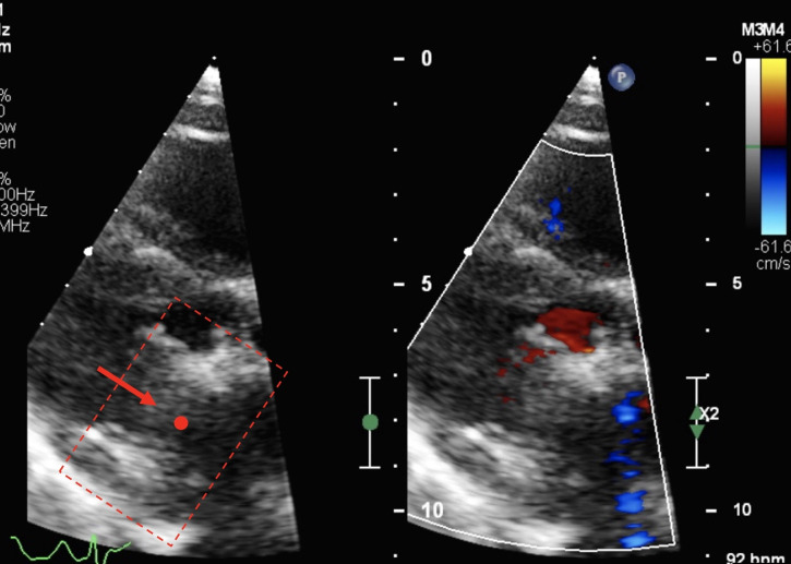
Cardiac disease after mediastinal radiotherapy can result in progressive valvular thickening and dystrophic calcification with ensuing leaflet restriction and dysfunction. This can ultimately manifest as valvular stenosis and/or regurgitation. We report a case of a 61-year-old woman with symptomatic severe aortic stenosis and severe mitral stenosis due to severe dystrophic calcification postmediastinal radiotherapy for lymphoma. She was deemed surgically inoperable due to dense, continuous calcification throughout the leaflets and annuli of both valves, aortomitral continuity, proximal coronary arteries and proximal porcelain aorta. She underwent simultaneous transcatheter aortic valve replacement and transcatheter mitral valve replacement with an excellent technical and clinical result at 7-month follow-up. We also describe the central role of multimodality three-dimensional transoesophageal echocardiography and multidetector cardiac CT imaging in assessing the severity of valve disease, characterising the nature of cardiac calcification and guiding decisions on surgical operability and transcatheter intervention.

Keywords: interventional cardiology; radiology (diagnostics); radiotherapy; surgical diagnostic tests; valvar diseases.


Conflict of interest statement

Competing interests: None declared.

Figures

Figure 1
Figure 1
Biplane TOE image (120 degree and 45 degree) demonstrating extensive calcification of mitral valve leaflets and annulus continuing into aortomitral continuity and aortic valve. TOE, transoesophageal echocardiography.
Figure 2
Figure 2
3D MPR demonstrating showing severe continuous calcification of the entirety of the mitral valve complex, through the aortomitral continuity into the aortic valve. 3D MPR, three-dimensional multiplanar reconstruction.
Figure 3
Figure 3
3D MPR demonstrating severe MS (left) and severe AS (right) planimetered at the leaflet tips. AS, aortic stenosis; 3D MPR, three-dimensional multiplanar reconstruction; MS, mitral stenosis.
Figure 4
Figure 4
3D multiplanar reconstruction (MPR) demonstrating continuous calcification from mitral valve leaflets (A–C, red dot) into mitral valve annulus (D), aortomitral continuity (E, green dot), aortic valve (F–G), aortic root (H) and ascending aorta (I).
Figure 5
Figure 5
MDCT 3D MPR demonstrating severe continuous calcification of the entirety of the mitral valve leaflets and annulus (red dot), through the aortomitral continuity (green arrow) into the aortic valve (yellow dot) and extending into the aortic root and ascending aorta (blue dot demonstrates circumferential calcification of porcelain aorta). Note that the top row centring on the mitral and aortic valves correlates exactly with the 3D TOE MPR (figure 3). 3D MPR, three-dimensional multiplanar reconstruction; MDCT, multidetector cardiac; TOE, transoesophageal echocardiography.
Figure 6
Figure 6
MDCT 3D volume-rendered reconstruction demonstrating severe continuous calcification of the entirety of the mitral valve leaflets and annulus into the aortic valve, aortic root and resultant porcelain ascending aorta. 3D, three dimensional; MDCT, multidetector cardiac.
Figure 7
Figure 7
TTE 7 months postprocedure with parasternal long-axis view demonstrating well-functioning TAVR with normal laminar antegrade flow through open valve (blue dot: TAVR orifice, blue arrow: direction of blood flow through TAVR, blue dotted box: TAVR valve). TAVR, transcatheter aortic valve replacement.
Figure 8
Figure 8
TTE 7 months postprocedure demonstrating well-functioning TMVR (red dot: TMVR orifice, red arrow: direction of blood flow through TMVR, red dotted box: TMVR valve).

References

    1. Gujral DM, Lloyd G, Bhattacharyya S. Radiation-induced valvular heart disease. Heart 2016;102:269–76. 10.1136/heartjnl-2015-308765 - DOI - PubMed
    1. Desai MY, Jellis CL, Kotecha R, et al. . Radiation-associated cardiac disease: a practical approach to diagnosis and management. JACC Cardiovasc Imaging 2018;11:1132–49. 10.1016/j.jcmg.2018.04.028 - DOI - PubMed
    1. Wyler von Ballmoos MC, Kalra A, Reardon MJ. Complexities of transcatheter mitral valve replacement (TMVR) and why it is not transcatheter aortic valve replacement (TAVR). Ann Cardiothorac Surg 2018;7:724–30. 10.21037/acs.2018.10.06 - DOI - PMC - PubMed
    1. Settergren M, De Palma R, Aortic CS. Combined severe aortic and mitral stenoses: an expanding potential for transcatheter therapies? JACC Cardiovasc Interv 2018;11:703–4. 10.1016/j.jcin.2018.01.269 - DOI - PubMed
    1. Kempfert J, Blumenstein JM, Borger MA, et al. . Minimally invasive off-pump valve-in-a-valve implantation: the atrial transcatheter approach for re-operative mitral valve replacement. Eur Heart J 2008;29:2382–7. 10.1093/eurheartj/ehn285 - DOI - PubMed

MeSH terms