Review
. 2021:42:519-550.
doi: 10.21775/cimb.042.519. Epub 2020 Dec 29.

Pathogenesis of Relapsing Fever

Affiliations
Review

Pathogenesis of Relapsing Fever

Job Lopez et al. Curr Issues Mol Biol. 2021.

Abstract

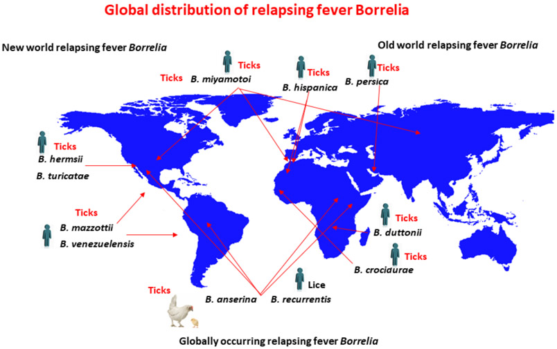
Relapsing fever (RF) is caused by several species of Borrelia; all, except two species, are transmitted to humans by soft (argasid) ticks. The species B. recurrentis is transmitted from one human to another by the body louse, while B. miyamotoi is vectored by hard-bodied ixodid tick species. RF Borrelia have several pathogenic features that facilitate invasion and dissemination in the infected host. In this article we discuss the dynamics of vector acquisition and subsequent transmission of RF Borrelia to their vertebrate hosts. We also review taxonomic challenges for RF Borrelia as new species have been isolated throughout the globe. Moreover, aspects of pathogenesis including symptomology, neurotropism, erythrocyte and platelet adhesion are discussed. We expound on RF Borrelia evasion strategies for innate and adaptive immunity, focusing on the most fundamental pathogenetic attributes, multiphasic antigenic variation. Lastly, we review new and emerging species of RF Borrelia and discuss future directions for this global disease.


Figures

Figure 1.
Figure 1.
Global distribution of RF Borrelia. Only certain representative RF Borrelia species are included in the figure. A more detailed description of currently defined species is found in (Barbour and Schwan, 2018; Talagrand-Reboul et al., 2018). Drawing was generated by Haitham Elbir. The global map was created using R software version 4.0.1
Figure 2.
Figure 2.
Antigenic variation and antibody response during RF borreliosis. Show are spirochete densities in the blood over time. As spirochetes replicate in the blood a predominant VMP variant emerges (pink line and spirochetes coated with pink proteins). The antibody response generated against this variant (pink dotted line) clears the population of spirochetes while a new population emerges (blue variant). Again, an antibody response is generated (blue dotted line) to clear the population, and yet another variant (green) emerges. The dynamics between antigenic variation and the host antibody response can continue for months. Drawing was generated using BioRender by Brittany A. Armstrong and Job Lopez.
Figure 3.
Figure 3.
Phase contrast micrograph of rosette of spirochetes and erythrocytes formed during experimental RF. Blood was examined four days after inoculation of an adult C3H/HeN mouse with Borrelia duttonii. Magnification 400X. Photo courtesy of Marie Andersson, University of Umeå.
Figure 4.
Figure 4.
Schematic representation of structure of a Vsp lipoprotein dimer of B. turicatae. The four major alpha-helical chains in each monomer are depicted by cylinders. The first and fourth alpha-helix (blue) in each Vsp are relatively conserved in sequence between different Vsp proteins of a given species (Dai et al., 2006; Restrepo et al., 1992). The second and third alpha-helical chain and the loops between all the chains are variable in sequence and are divided into variable regions (VR) 1 (red), 2 (green), 3 (yellow), and 4 (purple). The greatest variability is at the top or dome of the protein. Adapted from Lawson et al. (Lawson et al., 2006).
Figure 5.
Figure 5.
Three mechanisms for serotype switches by B. hermsii: allelic replacement, intramolecular DNA rearrangement, and in situ promoter activation. The different mechanisms are discussed in the text. P indicates the promoter and the location of the expression site near the right telomere of a linear plasmid; an overhead arrow shows whether the gene is active and the direction of transcription. vsp and vlp genes at the expression site location or in archival locations on the same (squiggly line background) or another linear plasmid (white background) are schematically represented by different patterns and grayscale values. The serotype of the cell, similarly indicated by pattern or grayscale, corresponds to vsp or vlp gene, at the expression site. In the second mechanism a deletion by direct repeats (small white horizontal bars) occurs and results in a non-replicative circle. (Courtesy of Alan Barbour, UC Irvine).
Figure 6.
Figure 6.
Organization of the expression plasmid (top) and one archival plasmid (bottom) in B. hermsii. Two examples of vsp or vlp genes are denoted by “X” and “Y” and different fill patterns. There is a single expression site on one plasmid, and silent variants of vsp and vlp genes are found on the same and other plasmids. The direction and extent of transcription of the duplicate gene at the expression site is indicated by the arrow. The UHS element at the expression site comprises 61 nt around the start codon of the variant gene (Barbour et al., 1991a; Dai et al., 2006; Kitten et al., 1993). Silent genes vary in the extent to which their UHS regions are identical to the expression site UHS; this is represented here by the relative length of the UHS block. There is a 214 nt noncoding DHS element downstream from the expression site and adjacent to the plasmid telomere and at various locations on the plasmids. The lengths of vsp/vlp gene examples and the UHS and DHS elements are not to scale. (Courtesy of Alan Barbour, UC Irvine).

References

    1. Aeschlimann A (1958). Développement embryonnaire d'Ornithodorus moubata (Murray) et transmission transovarienne de Borrelia duttoni. Acta Tropica 15, 15–64. - PubMed
    1. Alugupalli KR, Akira S, Lien E, and Leong JM (2007). MyD88- and Bruton's tyrosine kinase-mediated signals are essential for T cell-independent pathogen-specific IgM responses. J Immunol 178, 3740–3749. doi: 10.4049/jimmunol.178.6.3740 - DOI - PubMed
    1. Alugupalli KR, Gerstein RM, Chen J, Szomolanyi-Tsuda E, Woodland RT, and Leong JM (2003a). The resolution of relapsing fever borreliosis requires IgM and is concurrent with expansion of B1b lymphocytes. J Immunol 170, 3819–3827. doi:10.4049/jimmunol.170.7.3819 - DOI - PubMed
    1. Alugupalli KR, Leong JM, Woodland RT, Muramatsu M, Honjo T, and Gerstein RM (2004). B1b lymphocytes confer T cell-independent long-lasting immunity. Immunity 21, 379–390. doi:10.1016/j.immuni.2004.06.019 - DOI - PubMed
    1. Alugupalli KR, Michelson AD, Barnard MR, Robbins D, Coburn J, Baker EK, Ginsberg MH, Schwan TG, and Leong JM (2001). Platelet activation by a relapsing fever spirochaete results in enhanced bacterium-platelet interaction via integrin alphaIIbbeta3 activation. Mol Microbiol 39, 330–340. doi:10.1046/j.1365-2958.2001.02201.x - DOI - PubMed

LinkOut - more resources