. 2020 Dec 28;5(1):299.
doi: 10.1038/s41392-020-00438-7.

Severe acute respiratory syndrome coronavirus 2 (SARS-CoV-2) membrane (M) protein inhibits type I and III interferon production by targeting RIG-I/MDA-5 signaling

Affiliations

Severe acute respiratory syndrome coronavirus 2 (SARS-CoV-2) membrane (M) protein inhibits type I and III interferon production by targeting RIG-I/MDA-5 signaling

Yi Zheng et al. Signal Transduct Target Ther. .

Abstract

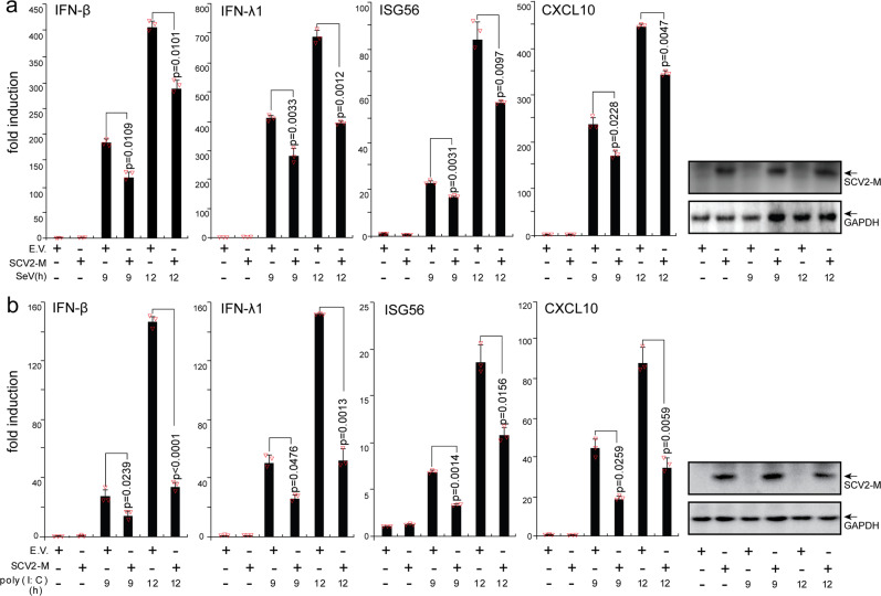
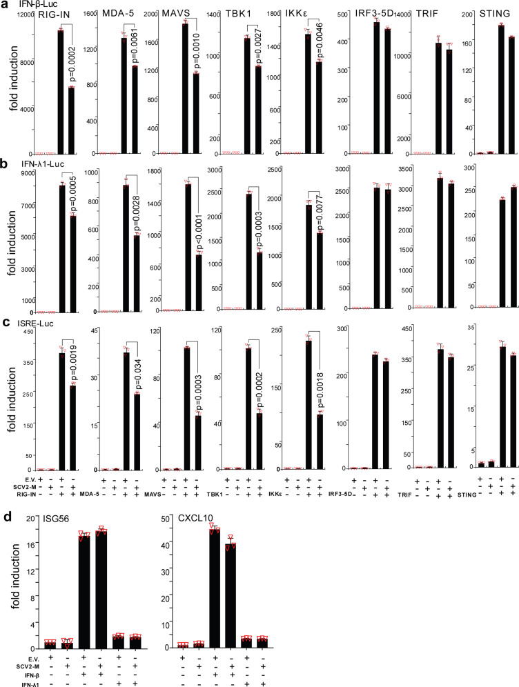
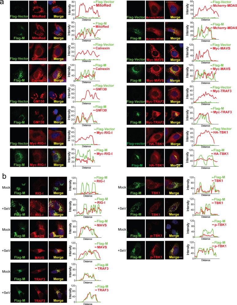
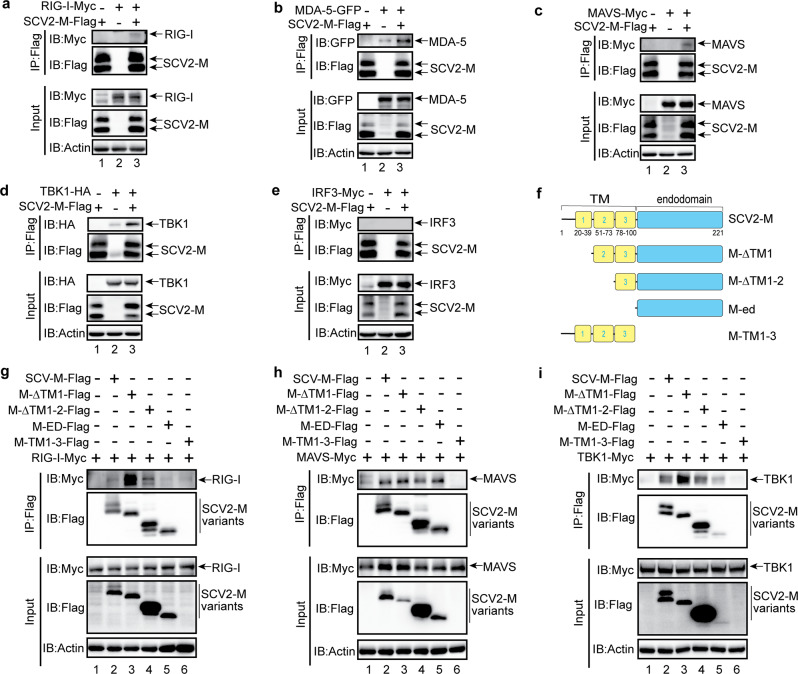
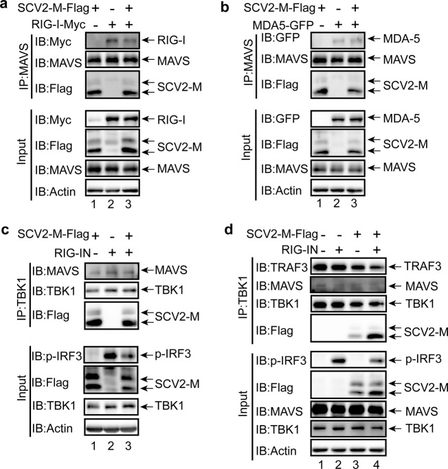
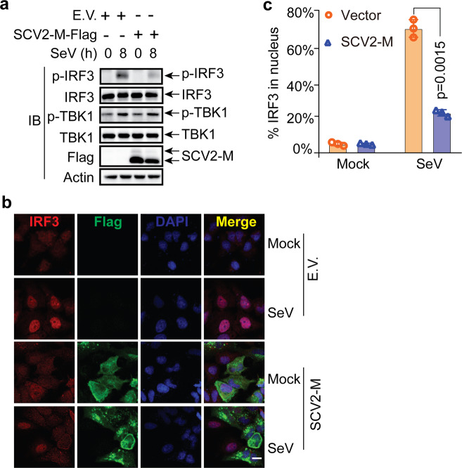
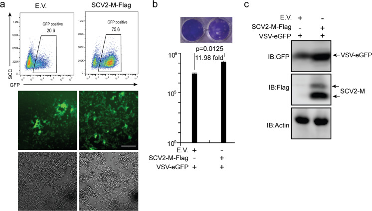
Coronavirus disease 2019 (COVID-19), caused by severe acute respiratory syndrome coronavirus 2 (SARS-CoV-2), has quickly spread worldwide and has affected more than 10 million individuals. A typical feature of COVID-19 is the suppression of type I and III interferon (IFN)-mediated antiviral immunity. However, the molecular mechanism by which SARS-CoV-2 evades antiviral immunity remains elusive. Here, we reported that the SARS-CoV-2 membrane (M) protein inhibits the production of type I and III IFNs induced by the cytosolic dsRNA-sensing pathway mediated by RIG-I/MDA-5-MAVS signaling. In addition, the SARS-CoV-2 M protein suppresses type I and III IFN induction stimulated by SeV infection or poly (I:C) transfection. Mechanistically, the SARS-CoV-2 M protein interacts with RIG-I, MAVS, and TBK1, thus preventing the formation of the multiprotein complex containing RIG-I, MAVS, TRAF3, and TBK1 and subsequently impeding the phosphorylation, nuclear translocation, and activation of IRF3. Consequently, ectopic expression of the SARS-CoV-2 M protein facilitates the replication of vesicular stomatitis virus. Taken together, these results indicate that the SARS-CoV-2 M protein antagonizes type I and III IFN production by targeting RIG-I/MDA-5 signaling, which subsequently attenuates antiviral immunity and enhances viral replication. This study provides insight into the interpretation of SARS-CoV-2-induced antiviral immune suppression and illuminates the pathogenic mechanism of COVID-19.


Conflict of interest statement

The authors declare no competing interests.

Figures

Fig. 1
Fig. 1
The SARS-CoV-2 M protein inhibits the induction of IFN-β, IFN-λ1, ISG56, and CXCL10 by SeV infection and poly (I:C) transfection. HEK293T cells cultured in 24-well plates (0.8–1 × 105 cells per well) were transfected with pcDNA6B empty vector (E.V., 500 ng) or the SCV2-M plasmid (500 ng). Twenty-four hours after transfection, cells were stimulated by SeV infection (a) or poly (I:C) transfection (b) as indicated, and at 9 and 12 h after stimulation, the cells were harvested for RNA extraction and subsequent RT-qPCR analysis. Three independent biological replicates were analyzed, the results of one representative experiment are shown, and the error bars indicate the SD values. Statistical significance is shown as indicated. SARS-CoV-2 M protein SCV2-M; hours h
Fig. 2
Fig. 2
The SARS-CoV-2 M protein suppresses the activation of the luciferase reporters of type I and III IFNs and ISGs. The pcDNA6B empty vector and the SARS-CoV-2 M protein plasmids (100 ng) were transfected with the indicated combinations of plasmids expressing RIG-IN (100 ng), MDA-5 (100 ng), TBK1 (100 ng), IKKε (100 ng), IRF3-5D (100 ng, an active form of IRF3), TRIF (100 ng, component of TLR3-TRIF pathway), or STING (100 ng, component of cGAS-STING pathway) into HEK293T cells cultured in 48-well plates (0.5 × 105 cells per well). The IFN-β-Luc (45 ng, the IFN-β luciferase reporter) (a), IFN-λ1-Luc (45 ng, the IFN-λ1 luciferase reporter) (b), or ISRE-Luc (45 ng, the IFN-stimulated response element luciferase reporter) (c) plasmids were also transfected to assess the activation of type I IFNs, type III IFNs, or ISGs, respectively. The pRL-TK (5 ng) was transfected into each well as an internal control. The pcDNA6B empty vector was used to normalize the total amount of transfected plasmid DNA. Dual‐luciferase assays were performed 36 h after transfection. d The pcDNA6B empty vector and the SARS-CoV-2 M protein plasmids (100 ng) were transfected into HEK293T cells as indicated. Twenty-four hours later, the cells were treated with recombinant human IFN-β (10 ng/mL) or IFN-λ1 (10 ng/mL). Three hours after simulation, the cells were harvested for RNA extraction and subsequent RT-qPCR analysis. Three independent biological replicates were analyzed, the results of one representative experiment are shown, and the error bars indicate the SD values. Statistical significance is shown as indicated. SARS-CoV-2 M protein SCV2-M
Fig. 3
Fig. 3
Subcellular localization of the SARS-CoV-2 M protein. HeLa cells seeded on coverslips in 12-well plates were transfected with the indicated plasmids. Twenty hours later, the cells were fixed, blocked, and then incubated with a rabbit anti-Flag antibody and a mouse antibody against the corresponding organelle marker or the indicated protein (a). HeLa cells in (b) were infected with SeV as indicated for 8 h before fixation, blocking and antibody incubation. Subsequently, the proteins were stained with a fluorescence-labeled secondary antibody. MDA-5 was visualized with a mCherry tag. (Right) Intensity profiles of SARS-CoV-2 M protein (Flag-M) and indicated proteins along the plotted lines, as analyzed by Image J line scan analysis. Nucleus were visualized with DAPI (blue). Confocal imaging results are representative of two independent experiments. Scale bar, 10 μm. MitoRed, mitochondria marker; Calnexin, ER marker; CM130, Golgi marker
Fig. 4
Fig. 4
The SARS-CoV-2 M protein interacts with RIG-I (a), MDA-5 (b), MAVS (c), and TBK1 (d) but not with IRF3 (e). f The truncated variants of SARS-CoV-2 M protein are depicted. g–i The interaction of RIG-I, MAVS, and TBK1 with the truncated variants of SARS-CoV-2 M protein. The HEK293T cells were transfected with the indicated plasmids for 24 h before coimmunoprecipitation with the anti-Flag magnetic beads. The pcDNA6B empty vector was used to balance the total amount of plasmid DNA in the transfection. The input and immunoprecipitates were immunoblotted with the indicated antibodies. Immunoblotting results are representative of two independent experiments. SARS-CoV-2 M protein, SCV2-M
Fig. 5
Fig. 5
The SARS-CoV-2 M protein prevents the formation of the RIG-I–MAVS–TRAF3–TBK1 multiprotein complex. The SARS-CoV-2 M protein inhibits the RIG-I–MAVS (a), MAVS–TBK1 (c), and TRAF3–TBK1 (d) but not MDA-5–MAVS (b) interactions. The HEK293T cells were transfected with the indicated plasmids for 24 h before coimmunoprecipitation with the antibodies against MAVS (a and b) or TBK1 (c and d). The pcDNA6B empty vector was used to balance the total amount of plasmid DNA in the transfection. The input and immunoprecipitates were immunoblotted with the indicated antibodies. Immunoblotting results are representative of two independent experiments. SARS-CoV-2 M protein, SCV2-M
Fig. 6
Fig. 6
The SARS-CoV-2 M protein suppresses the phosphorylation and nuclear translocation of IRF3. a The SARS-CoV-2 M protein affects the phosphorylation of IRF3 upon SeV infection. HeLa cells seeded in six-well plates were transfected with the empty vector or Flag-tagged SARS-CoV-2 M protein plasmids for 20 h before infection with SeV (50 HA/mL). At the indicated time points, the cells were harvested and processed for immunoblotting with the indicated antibodies. b The SARS-CoV-2 M protein prevents the nuclear translocation of IRF3. HeLa cells seeded on coverslips in 12-well plates were transfected with the Flag empty vector or Flag-tagged SARS-CoV-2 M protein plasmids for 20 h before infection with SeV. After infection for 8 h, the coverslips were removed and processed for immunofluorescence staining with the mouse anti-Flag antibody and a rabbit anti-IRF3 antibody. Scale bar, 10 μm. c Quantification of the percentage of IRF3 in the nucleus upon SeV infection. IRF3 molecules in the nuclei in 50 cells per group were counted, and percentages were calculated. Experiments were performed in triplicate and calculated as the means ± SD. Immunoblotting (a) and confocal imaging (b) results are representative of two independent experiments. Empty vector EV, SARS-CoV-2 M protein SCV2-M, hours h
Fig. 7
Fig. 7
The SARS-CoV-2 M protein facilitates viral replication. The HEK293T cells were transfected with plasmids as indicated. Twenty-four hours later, the cells were infected with VSV-eGFP (MOI = 0.001). Twelve hours after infection, GFP-positive cells were visualized and analyzed with flow cytometry (a), and the culture supernatant (20 h post-infection) was collected for plaque assays to measure the titer of extracellular VSV-eGFP (PFU/mL) (b). Confocal imaging and flow cytometry results are representative of two independent experiments. Scale bar, 50 μm. Three independent biological replicates were analyzed (a); the results of one representative experiment are shown, and the error bars indicate the SD value. The statistical significance is shown as indicated. c The replication of intracellular VSV-eGFP in the cell lysate (20 h post-infection) was determined by immunoblotting using an anti-GFP antibody. Empty vector EV; SARS-CoV-2 M protein SCV2-M

Similar articles

Cited by

References

    1. Wu F, et al. A new coronavirus associated with human respiratory disease in China. Nature. 2020;579:265–269. doi: 10.1038/s41586-020-2008-3. - DOI - PMC - PubMed
    1. Zhou P, et al. A pneumonia outbreak associated with a new coronavirus of probable bat origin. Nature. 2020;579:270–273. doi: 10.1038/s41586-020-2012-7. - DOI - PMC - PubMed
    1. Zhu N, et al. A novel coronavirus from patients with pneumonia in China, 2019. N. Engl. J. Med. 2020;382:727–733. doi: 10.1056/NEJMoa2001017. - DOI - PMC - PubMed
    1. Cui J, Li F, Shi ZL. Origin and evolution of pathogenic coronaviruses. Nat. Rev. Microbiol. 2019;17:181–192. doi: 10.1038/s41579-018-0118-9. - DOI - PMC - PubMed
    1. Kim D, et al. The architecture of SARS-CoV-2 transcriptome. Cell. 2020;181:914–921.e910. doi: 10.1016/j.cell.2020.04.011. - DOI - PMC - PubMed

Publication types

MeSH terms