. 2020 Dec 1;61(14):36.
doi: 10.1167/iovs.61.14.36.

Spectrum of Disease Severity in Patients With X-Linked Retinitis Pigmentosa Due to RPGR Mutations

Affiliations

Spectrum of Disease Severity in Patients With X-Linked Retinitis Pigmentosa Due to RPGR Mutations

Valentina Di Iorio et al. Invest Ophthalmol Vis Sci. .

Abstract

Purpose: The purpose of this study was to perform a detailed longitudinal phenotyping of X-linked retinitis pigmentosa (RP) caused by mutations in the RPGR gene during a long follow-up period.

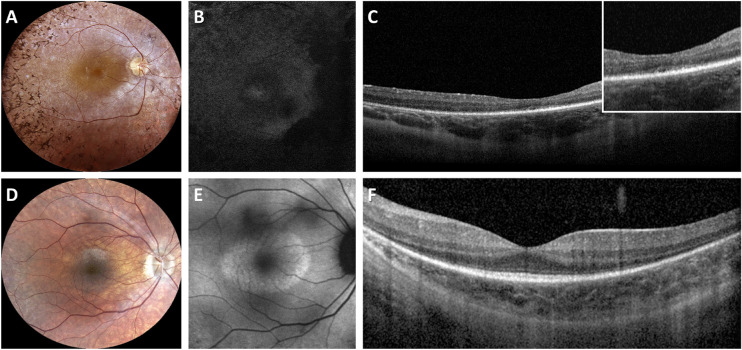
Methods: An Italian cohort of 48 male patients (from 31 unrelated families) with RPGR-associated RP was clinically assessed at a single center (mean follow-up = 6.5 years), including measurements of best-corrected visual acuity (BCVA), Goldmann visual field (GVF), optical coherence tomography (OCT), fundus autofluorescence (FAF), microperimetry, and full-field electroretinography (ERG).

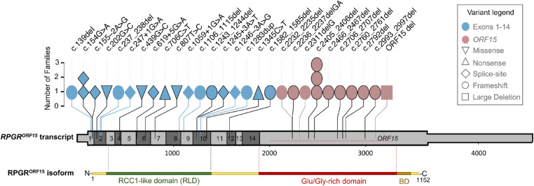
Results: Patients (29.6 ± 15.2 years) showed a mean BCVA of 0.6 ± 0.7 logMAR, mostly with myopic refraction (79.2%). Thirty patients (62.5%) presented a typical RP fundus, while the remaining sine pigmento RP. Over the follow-up, BCVA significantly declined at a mean rate of 0.025 logMAR/year. Typical RP and high myopia were associated with a significantly faster decline of BCVA. Blindness was driven primarily by GVF loss. ERG responses with a rod-cone pattern of dysfunction were detectable in patients (50%) that were significantly younger and more frequently presented sine pigmento RP. Thirteen patients (27.1%) had macular abnormalities without cystoid macular edema. Patients (50%) with a perimacular hyper-FAF ring were significantly younger, had a higher BCVA and a better-preserved ellipsoid zone band than those with markedly decreased FAF. Patients harboring pathogenic variants in exons 1 to 14 showed a milder phenotype compared to those with ORF15 mutations.

Conclusions: Our monocentric, longitudinal retrospective study revealed a spectrum disease progression in male patients with RPGR-associated RP. Slow disease progression correlated with sine pigmento RP, absence of high myopia, and mutations in RPGR exons 1 to 14.


Conflict of interest statement

Disclosure: V. Di Iorio, None; M. Karali, None; P. Melillo, None; F. Testa, None; R. Brunetti-Pierri, None; F. Musacchia, None; C. Condroyer, None; J. Neidhardt, None; I. Audo, None; C. Zeitz, None; S. Banfi, None; F. Simonelli, None

Figures

Figure 1.
Figure 1.
Multimodal imaging findings in two representative patients with RPGR-associated RP. (A) Fundus photograph of a 31-year-old patient with typical RP (ID no. 13; BCVA 0,3; NM_001034853: c.1245+3A>T) showing bone-spicule pigment deposits in the mid peripheral retina, optic disc pallor and RPE atrophy. (B) Fundus autofluorescence (FAF) image showing a widespread hypoautofluorescence. (C) Spectral domain OCT revealing RPE atrophy. The inset shows the disruption of the ellipsoid zone (EZ) band. (D) Fundus photograph of a 25-year-old patient with sine pigmento RP (ID no. 37; BCVA 0,7; NM_001034853: c.1059+1G>A) showing absence of bone-spicule-like pigment migration, optic disc pallor, and mild RPE atrophy. (E) FAF image showing a hyperautofluorescent ring around the central macula. (F) Spectral domain OCT showing central sparing of the EZ band that is attenuated toward the peripheral macula.
Figure 2.
Figure 2.
Survival curves of low vision and legal blindness (based on visual acuity and visual field) for patients with RPGR - associated RP. Kaplan–Meier plot showing the risk of developing low vision (green) and legal blindness, based on visual field (blue) or on visual acuity (red), with age. Survival analyses showed that legal blindness based on visual field is reached at a median age of 26.4 years (blue circle), low vision at 48.2 years (green circle), and legal blindness based on visual acuity at 51.3 years (red circle).
Figure 3.
Figure 3.
Genetic variants detected in RPGR. Schematic drawing of the RPGRORF15 transcript showing the position and frequency of the variants identified in this cohort. The untranslated regions of the transcript are depicted as a thinner bar. Each symbol designates a family whose variant position is reported on the top. The symbol color indicates whether the variant is found in exons 1 to 14 or in the ORF15 region. The symbol shape indicates the mutation type. The large deletion (ORF15 del) is shown with a horizontal dotted line within the terminal exon of RPGRORF15. Already reported variants are denoted with a black border. Protein domains of the retina-specific isoform RPGRORF15 are shown below the transcript to indicate the RCC1-like domain (green), the Glu/Gly-rich region (red), and the basic domain (BD; brown; not to scale).

References

    1. Hamel C. Retinitis pigmentosa. Orphanet J Rare Dis. 2006; 1: 40. - PMC - PubMed
    1. Chang S, Vaccarella L, Olatunji S, Cebulla C, Christoforidis J.. Diagnostic challenges in retinitis pigmentosa: genotypic multiplicity and phenotypic variability. Curr Genomics. 2011; 12: 267–275. - PMC - PubMed
    1. Branham K, Othman M, Brumm M, et al. .. Mutations in RPGR and RP2 account for 15% of males with simplex retinal degenerative disease. Invest Ophthalmol Vis Sci. 2012; 53: 8232–8237. - PMC - PubMed
    1. Lyraki R, Megaw R, Hurd T.. Disease mechanisms of X-linked retinitis pigmentosa due to RP2 and RPGR mutations. Biochem Soc Trans. 2016; 44: 1235–1244. - PubMed
    1. Tee JJ, Smith AJ, Hardcastle AJ, Michaelides M.. RPGR-associated retinopathy: clinical features, molecular genetics, animal models and therapeutic options. Br J Ophthalmol. 2016; 100: 1022–1027. - PubMed

Publication types