. 2021 Feb 8;6(3):e138385.
doi: 10.1172/jci.insight.138385.

Antineutrophil properties of natural gingerols in models of lupus

Affiliations

Antineutrophil properties of natural gingerols in models of lupus

Ramadan A Ali et al. JCI Insight. .

Abstract

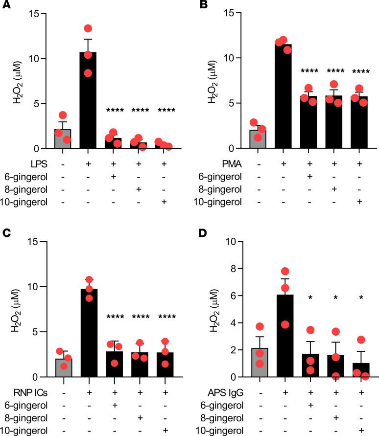
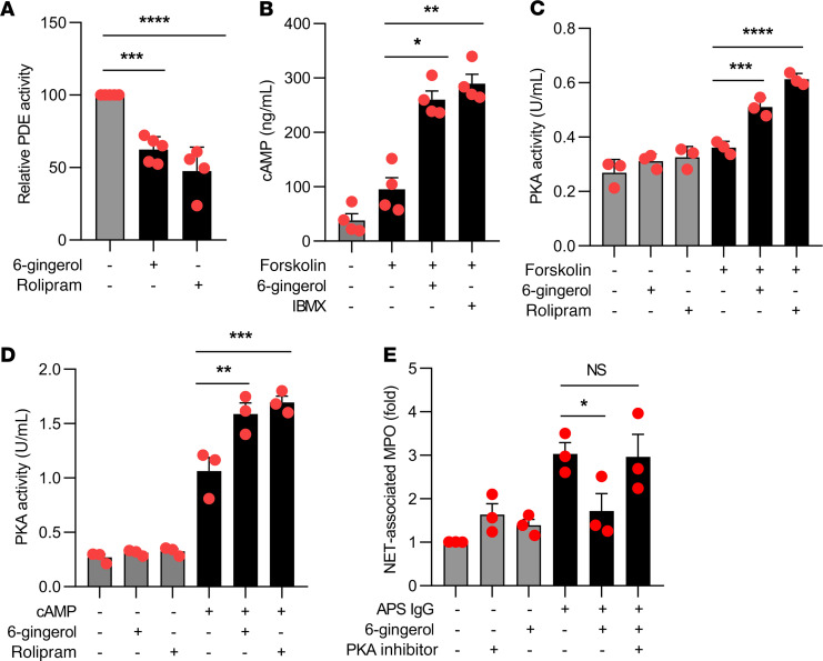
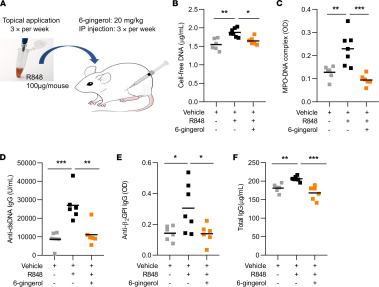
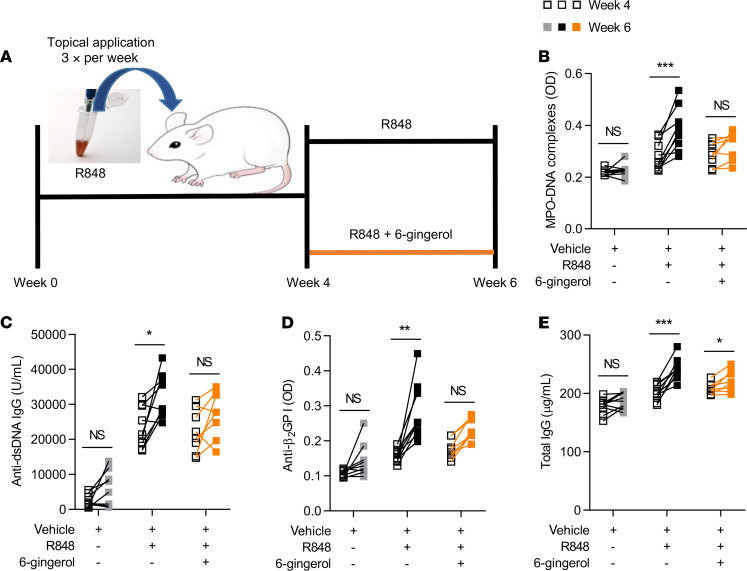
Ginger is known to have antiinflammatory and antioxidative effects and has traditionally been used as an herbal supplement in the treatment of various chronic diseases. Here, we report antineutrophil properties of 6-gingerol, the most abundant bioactive compound of ginger root, in models of lupus and antiphospholipid syndrome (APS). Specifically, we demonstrate that 6-gingerol attenuates neutrophil extracellular trap (NET) release in response to lupus- and APS-relevant stimuli through a mechanism that is at least partially dependent on inhibition of phosphodiesterases. At the same time, administration of 6-gingerol to mice reduces NET release in various models of lupus and APS, while also improving other disease-relevant endpoints, such as autoantibody formation and large-vein thrombosis. In summary, this study is the first to our knowledge to demonstrate a protective role for ginger-derived compounds in the context of lupus. Importantly, it provides a potential mechanism for these effects via phosphodiesterase inhibition and attenuation of neutrophil hyperactivity.

Keywords: Autoimmunity; Inflammation; Lupus; Neutrophils; Phosphodiesterases.


Conflict of interest statement

Conflict of interest: The authors have declared that no conflict of interest exists.

Figures

Figure 1
Figure 1. Gingerol suppresses NETosis in response to various stimuli.
Human neutrophils were isolated from healthy volunteers and then treated with various stimuli for 3 hours in the presence of different gingerol analogues. NETosis was quantified by measuring the enzymatic activity of nuclease-liberated myeloperoxidase (MPO). Dose response to LPS-mediated NETosis upon treatment with 6-gingerol (A), 8-gingerol (B), and 10-gingerol (C). NETosis in response to PMA (D), RNP ICs (E), and APS IgG (F) was quantified in the presence of 10 μM gingerol. NETosis was assessed by immunofluorescence microscopy (G). Neutrophils were treated with LPS, PMA, RNP ICs, or APS IgG in the presence or absence of 6-gingerol (10 μM). Blue, DNA; green, extracellular neutrophil elastase. Scale bar: 100 microns. For AF, mean and SEM are presented for n = 3 independent experiments; *P < 0.05, **P < 0.01, ****P < 0.0001 as compared with the 0 μM gingerol group by 1-way ANOVA corrected with Dunnett’s test.
Figure 2
Figure 2. Gingerols suppress ROS.
Human neutrophils were treated with various stimuli in the presence of different gingerol analogs for 1 hour. Hydrogen peroxide formation was measured by a colorimetric assay. Mean and SEM are presented for n = 3 independent experiments; *P < 0.05, ****P < 0.0001 as compared with the LPS-alone group (A), PMA-alone group (B), RNP ICs–alone group (C), or APS IgG–alone group (D) by 1-way ANOVA corrected with Dunnett’s test.
Figure 3
Figure 3. 6-Gingerol blocks PDE activity and raises cAMP levels.
Human neutrophils were treated with 6-gingerol. Some samples were additionally treated with forskolin, cAMP, and synthetic PDE4 inhibitors (rolipram and IBMX) as indicated. PDE activity (A), cAMP levels (B), and PKA activity (C and D) were measured with kits as described in Methods. In E, neutrophils were treated with APS IgG in the presence or absence of 6-gingerol and/or PKA inhibitor. NETosis was quantified by measuring the enzymatic activity of nuclease-liberated myeloperoxidase (MPO). Mean and SEM are presented for n = 3–4 independent experiments; *P < 0.05, **P < 0.01, ***P < 0.001, ****P < 0.0001 by 1-way ANOVA corrected with Dunnett’s test.
Figure 4
Figure 4. 6-Gingerol attenuates NET release and autoantibody formation in a lupus mouse model.
BALB/c mice were treated topically with TLR7 agonist (R848) or vehicle DMSO for 6 weeks (3 times per week). Some mice were additionally injected (i.p.) with 20 mg/kg 6-gingerol (3 times per week). Schematic of the TLR7 agonist–induced (R848) lupus model (A). NET levels in serum were assessed by measuring cell-free DNA (B) and MPO-DNA complexes (C). Anti–double-stranded DNA (anti-dsDNA) (D), anti–β-2 glycoprotein I (β2GPI) IgG (E), and total IgG (F) levels in serum were assessed by ELISA. Mean is presented as a horizontal line; *P < 0.05, **P < 0.01, ***P < 0.001 as compared with the R848-alone group by 1-way ANOVA corrected with Dunnett’s test.
Figure 5
Figure 5. Efficacy of 6-gingerol treatment after development of lupus phenotype in a lupus mouse model.
BALB/c mice were treated topically with TLR7 agonist (R848) or vehicle DMSO for 6 weeks (3 times per week). Starting at week 4 of treatment, some mice were additionally injected (i.p.) with 20 mg/kg 6-gingerol (3 times per week). Schematic of the TLR7 agonist–induced (R848) lupus model by week 4 followed by 6-gingerol treatment (A). NET levels in serum were assessed before and after 6-gingerol treatment by measuring MPO-DNA complexes (B). Anti–double-stranded DNA (anti-dsDNA) (C), anti–β-2 glycoprotein I (β2GPI) IgG (D), and total IgG (E) levels in serum were assessed by ELISA before and after 6-gingerol treatment. Mean is presented as a horizontal line; *P < 0.05, **P < 0.01, ***P < 0.001 by paired t test.
Figure 6
Figure 6. 6-Gingerol prevents aPL-mediated acceleration of venous thrombosis.
Schematic of the electrolytic model of venous thrombosis (A). Direct current results in the release of free radicals within the inferior vena cava, which activate endothelial cells and initiate a thrombogenic environment in the presence of constant blood flow. MPO-DNA complexes were measured in serum of mice treated with control IgG or APS IgG in the presence or absence of 6-gingerol (B). Thrombus formation was assessed at 24 hours. Thrombus length (C) and thrombus weight (D) were measured. Representative thrombi (E). **P < 0.01, ***P < 0.001, ****P < 0.0001 by 1-way ANOVA corrected with Dunnett’s test.
Figure 7
Figure 7. The synthetic PDE4 inhibitor rolipram suppresses APS-mediated NETosis and venous thrombosis.
Human neutrophils were stimulated with APS IgG for 3 hours. Some samples were additionally treated with the PDE4 inhibitor rolipram. NETosis was quantified by measuring the enzymatic activity of nuclease-liberated myeloperoxidase (MPO) (A). MPO-DNA complexes were assessed for control IgG- or APS IgG-treated mice in the presence or absence of rolipram (B). Thrombus formation was assessed at 24 hours. Thrombus length (C) and thrombus weight (D) were measured; **P < 0.01, ***P < 0.001, ****P < 0.0001 by 1-way ANOVA corrected with Dunnett’s test.
Figure 8
Figure 8. Kinetics of 6-gingerol in plasma and neutrophils following injection (i.p.) with 20 mg/kg 6-gingerol.
Male C57BL/6 mice were injected (i.p.) with 20 mg/kg 6-gingerol, and peripheral blood was collected after 0.5, 2, 4, and 24 hours. 6-Gingerol concentrations in plasma (A) and in neutrophils (B) were then quantitated. Values and error bars represent mean ± SEM, respectively.

Comment in

References

    1. Miyakis S, et al. International consensus statement on an update of the classification criteria for definite antiphospholipid syndrome (APS) J Thromb Haemost. 2006;4(2):295–306. doi: 10.1111/j.1538-7836.2006.01753.x. - DOI - PubMed
    1. Bertolaccini ML, et al. 14th International congress on antiphospholipid antibodies task force. Report on antiphospholipid syndrome laboratory diagnostics and trends. Autoimmun Rev. 2014;13(9):917–930. doi: 10.1016/j.autrev.2014.05.001. - DOI - PubMed
    1. Gómez-Puerta JA, Cervera R. Diagnosis and classification of the antiphospholipid syndrome. J Autoimmun. 2014;48–49:20–25:.:S0896–8411(14)00008–0. doi: 10.1016/j.jaut.2014.01.006. - DOI - PubMed
    1. Cervera R, et al. Antiphospholipid syndrome: clinical and immunologic manifestations and patterns of disease expression in a cohort of 1,000 patients. Arthritis Rheum. 2002;46(4):1019–1027. doi: 10.1002/art.10187. - DOI - PubMed
    1. Brinkmann V, et al. Neutrophil extracellular traps kill bacteria. Science. 2004;303(5663):1532–1535. doi: 10.1126/science.1092385. - DOI - PubMed

Publication types

MeSH terms