. 2020 Dec;14(6):910-920.
doi: 10.31616/asj.2020.0593. Epub 2020 Dec 22.

Diagnostic Modality in Spine Disease: A Review

Affiliations

Diagnostic Modality in Spine Disease: A Review

Gang-Un Kim et al. Asian Spine J. 2020 Dec.

Abstract

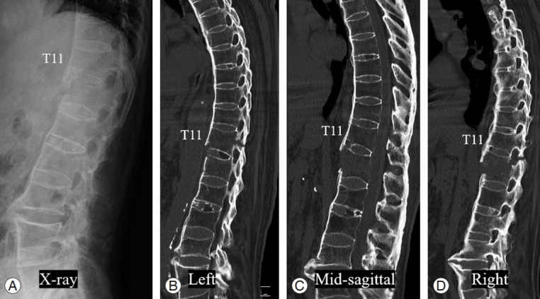
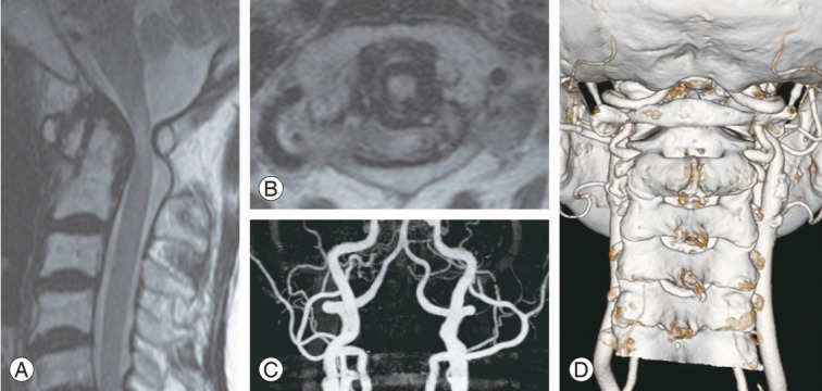
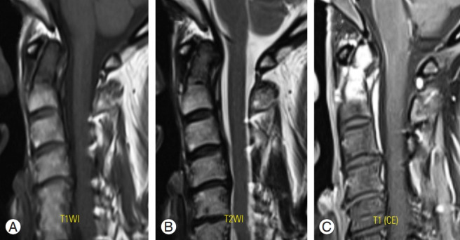
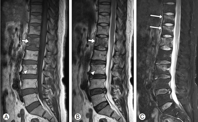
Spine diseases are common and exhibit several causes, including degeneration, trauma, congenital issues, and other specific factors. Most people experience a variety of symptoms of spine diseases during their lifetime that are occasionally managed with conservative or surgical treatments. Accurate diagnosis of the spine pathology is essential for the appropriate management of spine disease, and various imaging modalities can be used for the diagnosis, including radiography, computed tomography (CT), magnetic resonance imaging (MRI), and other studies such as EOS, bone scan, single photon emission CT/CT, and electrophysiologic test. Patient (or case)-specific selection of the diagnostic modality is crucial; thus, we should be aware of basic information and approaches of the diagnostic modalities. In this review, we discuss in detail, about diagnostic modalities (radiography, CT, MRI, electrophysiologic study, and others) that are widely used for spine disease.

Keywords: Computed tomography; Diagnostic imaging; Electrophysiologic study; Magnetic resonance imaging; Radiography; Spinal diseases.


Conflict of interest statement

No potential conflict of interest relevant to this article was reported.

Figures

Fig. 1.
Fig. 1.
Lateral radiograph (A) and sagittal CT images (B–D) of the thoracolumbar spine showing full ankylosis of the spine, suspiciously ankylosing spondylitis, and multiple fractures at T10–12 levels with traumatic subluxation. The fracture site, pattern, and extent can be detected more clearly at CT images than those at lateral radiograph. CT, computed tomography.
Fig. 2.
Fig. 2.
(A, B) Cervical spine magnetic resonance imaging showing posteriorly subluxation of C2 odontoid process and signal change at the spinal cord at C1–2 level. (C, D) Three-dimensional reconstructed computed tomography images at C1–2 level showing bony pathologies as well as surrounding structures (especially, course, proximity, and other abnormalities of vertebral artery).
Fig. 3.
Fig. 3.
(A–C) Metastatic carcinoma with pathologic fracture on C2 odontoid process. CE T1-weighted MRI (C) showing increased signal intensity of the lesion at C2 odontoid process, that makes detection of the lesion more obviously in comparison with other sequences of images on MRI (A, B). CE, contrast enhanced; MRI, magnetic resonance imaging.
Fig. 4.
Fig. 4.
Sagittal T1- (A) and T2- (B) weighted magnetic resonance imaging of the lumbar spine showing definite signal change in L1 vertebral body (short arrows) and subtle marrow edema at the L3 vertebra (arrowheads). Additional T2 sagittal image with fat suppression technique showing the previously identified signal changes of L1 and L3 clearer, (C) while the occult fractures of the T11 and T12 vertebrae are clearly revealed due to the definite contrast of marrow edema (long arrows).
Fig. 5.
Fig. 5.
MRI and EMG of a 46-male patients having right lumbar radicular pain. Both examinations were conducted 1 month after the symptom onset. (A, B) T2-weighted MRIs at L4–5 disc level showed right central protrusion type disc herniation. (C) Positive sharp waves are manifested on right lumbar paraspinals and the muscles (right tensor fascia latae, tibialis anterior, and peroneus longus) innervated by right L5 nerve root, which is indicative finding of right L5 radiculopathy. MRI, magnetic resonance imaging; EMG, electromyography; Fib., fibrillation; Insert. ac., insertional activity; Bizz, bizzar potential; NMU, normal motor unit; LMU, large motor unit; Long po., long duration polyphasic potential; Short po., short duration polyphasic potential; Interf. pat., interference pattern.

Similar articles

Cited by

References

    1. Bansal GJ. Digital radiography. A comparison with modern conventional imaging. Postgrad Med J. 2006;82:425–8. - PMC - PubMed
    1. Leone A, Guglielmi G, Cassar-Pullicino VN, Bonomo L. Lumbar intervertebral instability: a review. Radiology. 2007;245:62–77. - PubMed
    1. Jahnke RW, Hart BL. Cervical stenosis, spondylosis, and herniated disc disease. Radiol Clin North Am. 1991;29:777–91. - PubMed
    1. Landman JA, Hoffman JC, Jr, Braun IF, Barrow DL. Value of computed tomographic myelography in the recognition of cervical herniated disk. AJNR Am J Neuroradiol. 1984;5:391–4. - PMC - PubMed
    1. Simon JE, Lukin RR. Diskogenic disease of the cervical spine. Semin Roentgenol. 1988;23:118–24. - PubMed

LinkOut - more resources