Review
. 2021 Mar 24;121(6):3412-3463.
doi: 10.1021/acs.chemrev.0c01010. Epub 2020 Dec 29.

β-Lactams against the Fortress of the Gram-Positive Staphylococcus aureus Bacterium

Affiliations
Review

β-Lactams against the Fortress of the Gram-Positive Staphylococcus aureus Bacterium

Jed F Fisher et al. Chem Rev. .

Abstract

The biological diversity of the unicellular bacteria-whether assessed by shape, food, metabolism, or ecological niche-surely rivals (if not exceeds) that of the multicellular eukaryotes. The relationship between bacteria whose ecological niche is the eukaryote, and the eukaryote, is often symbiosis or stasis. Some bacteria, however, seek advantage in this relationship. One of the most successful-to the disadvantage of the eukaryote-is the small (less than 1 μm diameter) and nearly spherical Staphylococcus aureus bacterium. For decades, successful clinical control of its infection has been accomplished using β-lactam antibiotics such as the penicillins and the cephalosporins. Over these same decades S. aureus has perfected resistance mechanisms against these antibiotics, which are then countered by new generations of β-lactam structure. This review addresses the current breadth of biochemical and microbiological efforts to preserve the future of the β-lactam antibiotics through a better understanding of how S. aureus protects the enzyme targets of the β-lactams, the penicillin-binding proteins. The penicillin-binding proteins are essential enzyme catalysts for the biosynthesis of the cell wall, and understanding how this cell wall is integrated into the protective cell envelope of the bacterium may identify new antibacterials and new adjuvants that preserve the efficacy of the β-lactams.


Conflict of interest statement

The authors declare no competing financial interest.

Figures
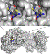
Figure 1.
Figure 1.
Top, the stereoview of S. aureus PBP3 acylated within the active site by the cephalosporin cefotaxime (PDB 3VSL). Activation of the active-site serine nucleophile (Ser392) is accomplished by a lysine general base. The perspective shown in Chart 2 for the cefotaxime-derived acyl-enzyme of PBP3 corresponds to this stereoview. The thiazolamine segment is in the foreground. The carbonyl of the acyl-enzyme is in the background. The nucleophilic oxygen of the serine is not visible (hidden behind the protein). Bottom, structure of the PBP3 cefotaxime-derived acyl-enzyme represented as a solvent-accessible surface with the bound antibiotic depicted space-filled and color-coded by atom types (blue for nitrogen, red for oxygen, yellow for sulfur, and gray for carbon). In this perspective the transpeptidase active site (to the left), here occupied by this acyl-enzyme, projects into the inner wall zone. The membrane-binding segment of PBP3 is not shown. Its location would be to the right of the protein.
Figure 2.
Figure 2.
This figure serves as an organizational guide to the three key structural entities of the Gram-positive cell envelope and thus gives context to many of the topics within this review. The horizontal center of the figure is the lipid bilayer of the single membrane of the Gram-positive bacterium. Above this membrane is the inner wall zone, above which the peptidoglycan cell wall (peptidoglycan synthesis by Lipid II polymerization and WTA glycopolymer attachment) is assembled. Within this membrane are (from left to right) the integral membrane transporters for the lipoteichoic acids (LTAs), for Lipid II, and for the wall teichoic acids (WTAs). Within the inner leaflet of this membrane are the membrane enzymes of the final biosynthetic steps of the lipid anchor of the lipoteichoic acids, of Lipid II, and of the wall teichoic acids. Lipid II and the WTAs share the common membrane carrier, undecaprenyl phosphate (Und-P). After their translocation this carrier is released, as the diphosphate, in the outer leaflet of the membrane. Efficient recycling of the Und-P carrier (not illustrated in this figure) is critical to balancing Lipid II and WTA availability. The combination of the lipid segment of the LTAs within the outer leaflet of the membrane, and the interdigitation of the glycopolymer of the LTA into the cell wall, conjoin the two and thus are essential to the structural integrity of the overall cell envelope. Covalent WTA attachment to the peptidoglycan creates a formidable exterior polymeric barrier for controlling solute access to the bacterium.
Figure 3.
Figure 3.
Cartoon schematic of the dividing S. aureus coccus. The gray spherical shell is the bacterial membrane. The turquoise spherical shell is the peptidoglycan. (A) The near-spherical coccus. (B) Midcell formation of a Z-ring (dashed-yellow circle) by inter alia GTP-dependent polymerization of the FtsZ protein. (C) Synthesis of new peptidoglycan (red) where the Z-ring is in contact with the old peptidoglycan, as a prelude for the invagination process of the cell envelope to enable cell division. The red peptidoglycan appears ultimately on the surface of the daughter cells as surface ribs (bottom left panel). These ribs (from previous cell division) are present in the bacterium of panels (A) and (B) but are not shown. (D) Progressive Z-ring constriction guides the synthesis of the septal peptidoglycan (dark-blue) built upon the red “rib” peptidoglycan. The blue peptidoglycan grows inward in a concentric motion of a leading edge, behind which the leading-edge peptidoglycan is progressively “thickened”. The different red-blue peptidoglycan coloration reflects both that different PBPs are used for the synthesis of two and the likely possibility that the polymeric structure of the two peptidoglycans is different. The white line centered in the blue peptidoglycan indicates a structural gap (of unknown structure or separation nature) created in the inward-growing peptidoglycan. (E) Septum formation is completed as an annulus fusion followed by (F) completion of the septal peptidoglycan. (G) Controlled degradation of the peptidoglycan external to red rib, and within the gap of the septum, prepares the cells for their final separation. This separation is driven by the internal osmotic pressure of the cells. The “popping” transition to give initially two hemispherically shaped daughter bacteria occurs on a millisecond time scale. Structural reshaping of the hemispherical bacteria to the near-spherical bacteria of the panel is likewise fast. Following division, the blue peptidoglycan is remodeled to give the uniformity of polymeric structure as indicated by the turquoise coloration of panel (A).
Figure 4.
Figure 4.
Peptidoglycan biosynthesis in the methicillin-susceptible S. aureus is accomplished by four PBP enzymes (PBPs 1–4). Although there is functional redundancy within the four and only PBP1 and PBP2 are essential, the pathogenic S. aureus bacterium requires all four PBP activities. Current mechanistic understanding suggests that synthesis of the red “rib” peptidoglycan (see Figure 3) is a primary task of PBP3; synthesis of the leading-edge septal peptidoglycan by the progressive concentric motion of the divisome is the primary task of PBP1 within the divisome complex; and the task of thickening the peptidoglycan toward structural strength, upon the leading-edge peptidoglycan, is the task of PBP2. PBP4 engages in the remodeling of the septal peptidoglycan and the wall peptidoglycan. In methicillin-resistant S. aureus, the essential transpeptidase-catalyzed cross-linking function of PBP2 is compromised by inactivation by the clinically achieved concentrations of the β-lactam antibiotics. Acquisition by these bacteria of the mec gene enables expression of a fifth PBP, that of PBP2a, that functions in complex with PBP2 to complete septal peptidoglycan synthesis. The transglycosylase activity of PBP2 coordinates with the transpeptidase activity of PBP2a for this completion. As inactivation of PBP2a requires higher β-lactam concentrations than can be achieved with almost all β-lactams, the PBP2·PBP2a pair continues to function, and the MRSA bacterium shows β-lactam resistance.
Figure 5.
Figure 5.
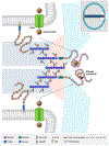
Cross-section cartoon perspective of the MRSA S. aureus cell envelope. This cartoon complements the structures shown in Scheme 2. This cartoon is suggestive of the structural organization of the envelope and is not intended to indicate a realism for that organization. Here the multiprotein, multienzyme divisome complex is represented by the integral membrane “flippase” MurJ (magenta) that delivers Lipid II to a PBP2 homodimer (monomers are colored in yellow and orange) in respective complex with two PBP2a enzymes (lime-green). The BlaZ β-lactamase resistance enzyme (light-purple) is a lipoprotein of the outer leaflet of the membrane. The membrane-anchored and structurally essential LTA molecules (dark-blue) interconnect the membrane (sky-blue) to the peptidoglycan. The molecular basis for the interaction between the LTAs and the peptidoglycan is not known. The LTAs do not project to the surface of the bacterium. The surface of the bacterium comprises the WTA molecules (purple) covalently attached to the peptidoglycan polymer (sea-green). The forest-green shadowing shown for the peptidoglycan indicates that the peptidoglycan is not a uniform polymer but has gaps and cavities. The density of both the LTAs and WTAs with respect to the peptidoglycan is greater than is suggested by the cartoon.
Figure 6.
Figure 6.
Schematic for the activation and turnover of BlaR. Antibiotic recognition on the cell surface by BlaR (left panel) leads to activation of its zinc-protease domain at the inner membrane-cytoplasm interface of this transmembrane protein. This protease activity degrades BlaI. As a result of the loss of BlaI the antibiotic-resistance genes of its operon, including that for BlaR1 itself, are derepressed. BlaR1 eventually experiences fragmentation at two sites, with cleavage at one shedding the sensor domain (BlaRS) from the membrane (right panel). This model of the BlaR protein is based on the corresponding model for the MecR protein as proposed by Belluzo et al.
Figure 7.
Figure 7.
(A) X-ray structure of the S. aureus PBP2a shown as a light gray solvent-accessible surface with a synthetic peptidoglycan fragment, depicted in space-filled presentation (carbons in dark gray, oxygens in red, and nitrogens in blue), bound to the allosteric site. (B) Stereoview of the allosteric site with the bound peptidoglycan and (C) of the unoccupied active site. The active site is approximately 60 Å distant from the allosteric site. The structural changes in the allosteric transformation that controls substrate access to the active-site serine, spanning the two sites, is understood by crystallographic evidence, computational simulations, and kinetic data.
Figure 8.
Figure 8.
Stereoview of the allosteric signal propagation in S. aureus PBP2a. Binding of the peptidoglycan (black structure at the allosteric site (between Lobe-1 and Lobe-2) propagates a network of salt-bridge interactions extending between the allosteric and catalytic domains (the transpeptidase active site is at the top of the enzyme). The seven salt-bridge interactions seen by crystallography are identified with arrowheads. The catalytic serine (yellow at 12 o’clock) and the acidic (red) and basic (blue) residues of the salt-bridge interactions are shown as spheres. Peptidoglycan (or small molecule) binding at the allosteric site stimulates a domino motion from the allosteric site (intersection of Lobe-1 and Lobe-2), through Lobe-3, and onto the β3–β4 loop that controls access to the active site.
Figure 9.
Figure 9.
Suggested integration of the structural components of the S. aureus cell envelope with respect to spatial control of the Atl autolysin in S. aureus cell division. The structural components are rendered in cartoon form and placed with reference to Panel G of Figure 3 (duplicated as the top right inset). The structural components are (7 o’clock to 3 o’clock) the LTA (decorated with d-Ala residues), the peptidoglycan (bifurcated to indicate growth of the dual septa of the daughter cells), and nascent WTA at the septal perimeter. The icons used for the saccharides follow glycan icon nomenclature (Glc, blue circle; GlcNAc, blue square; ManNAc, green square; MurNAc, purple hexagon). Nascent WTA is not decorated with d-Ala residues. The Atl pro-bifunctional autolysin enzyme, represented by yin and yang (light brown/dark brown) circle symbol, is transported to engage the nascent WTA either through or in coordination with the TarGH transporter (the arrows of the figure are meant to represent either possibility). Atl is held in place by electrostatic interaction with the nascent WTA. The mature WTA found elsewhere on the cell envelope is suggested to be decorated by d-Ala residues (by transacylation of the d-Ala residues of the LTA) and thus incapable of binding Atl. Accordingly, Atl is held to the septal perimeter. Atl activation is tightly regulated (by an unknown mechanism) to the final stage of cytokinesis. Based on observations made with S. pneumoniae, inactivation of PBPs by the β-lactams disrupts this regulation, leading to premature activation of Atl autolysin and disregulated peptidoglycan degradation. This degradation is suggested as the culminating event of the bactericidal mechanism of the β-lactams.
Chart 1.
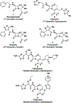
Chart 1.. Six Representative Structures of the β-Lactams Used in S. aureus Chemotherapya
aBenzylpenicillin 1 is a first-generation penicillin that lost quickly its clinical efficacy due to the acquisition by S. aureus of an enzyme, the BlaZ β-lactamase, which deactivated the penicillin by catalytic hydrolysis of its β-lactam ring to give the inactive β-amino acid metabolite. Cefazolin 2 is a first-generation cephalosporin that is a poor BlaZ substrate and thus is active against methicillin-susceptible S. aureus (MSSA). Oxacillin 3 and flucloxacillin 4 are second-generation penicillins of the methicillin class. They are poor BlaZ substrates and are still used in MSSA therapy. Ceftobiprole 5 and Ceftaroline 6 are the newest-generation cephalosporins with both Gram-positive and Gram-negative efficacy. In particular with respect to S. aureus, both structures have an enhanced ability to be recognized by and to inactivate the resistance penicillin-binding protein PBP2a of methicillin-resistant S. aureus (MRSA). Both drugs are used clinically as prodrug formulations.
Chart 2.
Chart 2.. The β-Lactams as Structural Mimetics of the d-Ala-d-Ala Stem Dipeptide Terminus of the Peptidoglycan
The A structures compare (left) the R-d-Ala-d-Ala dipeptide terminus of the stem peptide of the peptidoglycan to the structure (right) of a penicillin. The red color identifies the structure commonality as proposed by Tipper and Strominger. The left of the B structures is that of the cephalosporin cefotaxime. To its right is the acyl-enzyme structure of a PBP inactivated by cefotaxime. The mechanism of the inactivation is ring-opening of the β-lactam by the active-site serine nucleophile to give the stable acyl-enzyme. This acyl-enzyme is stable as it is unreactive for acyl-transfer. In normal PBP catalysis, the acyl moiety of a peptidoglycan-derived acyl-enzyme is transferred, as a crosslinking reaction, to the terminal amine of the bridge peptide of an adjacent peptidoglycan strand. The correlation between the irreversible incorporation of penicillins into the bacterial PBPs, and the bactericidal mechanism of the penicillins, by Strominger was a milestone both for mechanistic enzymology and for the determination of antibiotic mechanism.
Chart 3.
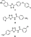
Chart 3.
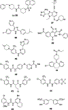
MRSA-Acting Oxadiazole Structures
Chart 4.
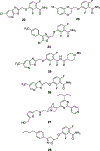
Chart 4.
MRSA-Acting Quinazolin-4-one Structures
Chart 5.
Chart 5.
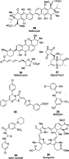
MRSA-Acting ClpP Inhibitors
Chart 6.
Chart 6.
MRSA-Acting FtsZ Inhibitors
Chart 7.
Chart 7.
MRSA-Acting Two-Component Kinase Inhibitors
Chart 8.
Chart 8.
MRSA-Acting Serine–Threonine Kinase Inhibitors
Scheme 1.
Scheme 1.. Mechanism of the β-Lactams Is PBP Inactivation by the Formation of a Stable Acyl-Enzyme Derived from the β-Lactama
aThis scheme provides spare kinetic summaries for PBP turnover of substrates and inactivation by β-lactams. The upper kinetic equation is substrate turnover. The PBP recognizes the R-d-Ala-d-Ala terminus of the peptidoglycan stem (see Scheme 2). From this Michaelis complex, an active-site lysine catalyzes opening of the β-lactam ring by a nucleophilic serine to give an acyl-enzyme intermediate. The PBP family divides between PBPs that catalyze peptidoglycan polymerization and peptidoglycan remodeling. In S. aureus the polymerizing PBPs transfer the acyl moiety, achieving a crosslinking reaction, to the amine of the terminal glycine residue of the bridge peptide of an adjacent peptidoglycan strand. In this scheme the bridge acyl-acceptor is abbreviated as R′-Gly. Note that the use of R′-Gly is not general, as different bacteria have different bridge structures. This kinetic sequence is contrasted with PBP inactivation by β-lactams. Here, the β-lactam is recognized as an R-d-Ala-d-Ala structural mimetic, and the active-site serine is acylated efficiently (lower kinetic equation). In contrast to PBP turnover, where there is departure of the terminal d-Ala as a leaving group, no leaving group departs upon β-lactam acylation of the active site serine. As a consequence, the β-lactam-derived acyl-enzyme (representative structure given in Chart 2) is incompetent for acyl-transfer. It is stable for multiple hours, far too long to sustain viability to the bacterium. The structural basis for the stability of the β-lactam-derived acyl-enzyme is steric interference with the acyl-acceptor (R′-Gly in polymerization reaction of S. aureus).,,
Scheme 2.
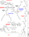
Scheme 2.. Principle Structures of the S. aureus Cell Envelopea
aThe cell envelope surrounds the cytoplasm of the bacterium in the following order: membrane (adjacent to the cytoplasm: here showing only the outer leaflet and with abbreviated acyl structures for the diacylglycerol); the inner-wall zone (contains many of the enzymes used in cell-envelope creation, not shown here); a wall teichoic acid (WTA, top left) attached covalently to the polymeric peptidoglycan (top left, below the wall teichoic acid). The wall teichoic acid–peptidoglycan is the surface structure of some pathogenic S. aureus strains. Many other S. aureus strains have polysaccharides (not shown) attached to the peptidoglycan. The lipoteichoic acid (LTA, right structure) extends from the membrane through the inner-wall zone and intercalates the peptidoglycan. LTAs are essential to the structural integrity of the envelope. Lipid II (bottom left) is assembled in the cytoplasm and translocated from the inner leaflet of the membrane to the outer leaflet of the membrane, with its disaccharide glycopeptide segment projecting into the inner-wall zone. Lipid II is the membrane-bound biosynthetic entity assembled into the peptidoglycan polymer. The Lipid II structure is parsed into four segments: an undecaprenol diphosphate membrane lipid, the NAG-NAM disaccharide, a pentapeptide stem whose last two amino acids are d-Ala-d-Ala, and a pentaglycine bridge attached to the ε-amine of the third amino acid (l-Lys) of the stem. Above the pentaglycine bridge of Lipid II is a nascent peptidoglycan strand (shown as a tetrasaccharide, formed from a transglycosylation reaction using Lipid II as the glycosyl donor adding to the terminal GlcNAc saccharide of a nascent peptidoglycan strand) that has been cross-linked (bridge-stem-bridge) to a second peptidoglycan strand. The dashed red oval to the left shows the functional group resulting from the cross-linking: and amide formed from the terminal glycine of the bridge to the carbonyl of the fourth amino acid (the penultimate d-Ala) of the stem. The second dashed red oval (top center) shows the reaction that forms this amide. The amine of the terminal glycine adds to the carbonyl of the (fourth amino acid of the stem) d-Ala, displacing the terminal d-Ala as the leaving group. This reaction is catalyzed by the Penicillin Binding Protein (PBP) enzymes, by a sequence of acyl-transfer to the active-site serine of the PBP, followed by acyl-transfer from this serine acyl-enzyme to the terminal amine of the Gly5 bridge.

References

    1. David MZ The importance of Staphylococcus aureus genotypes in outcomes and complications of bacteremia. Clin. Infect. Dis 2019, 69, 1878–1880. - PubMed
    1. Souli M; Ruffin F; Choi SH; Park LP; Gao S; Lent NC; Sharma-Kuinkel BK; Thaden JT; Maskarinec SA; Wanda L; et al. Changing characteristics of Staphylococcus aureus bacteremia: results from a 21-year, prospective, longitudinal study. Clin. Infect. Dis 2019, 69, 1868–1877. - PMC - PubMed
    1. Mulani MS; Kamble EE; Kumkar SN; Tawre MS; Pardesi KR Emerging strategies to combat ESKAPE pathogens in the era of antimicrobial resistance: A review. Front. Microbiol 2019, 10, 539. - PMC - PubMed
    1. Rello J; Eshwara VK; Lagunes L; Alves J; Wunderink RG; Conway-Morris A; Rojas JN; Alp E; Zhang Z A global priority list of the TOp TEn resistant Microorganisms (TOTEM) study at intensive care: a prioritization exercise based on multi-criteria decision analysis. Eur. J. Clin. Microbiol. Infect. Dis 2019, 38, 319–323. - PubMed
    1. Talbot GH; Jezek A; Murray BE; Jones RN; Ebright RH; Nau GJ; Rodvold KA; Newland JG; Boucher HW The Infectious Diseases Society of America’s 10 × ‘20 initiative (ten new systemic antibacterial agents FDA-approved by 2020): Is 20 × ‘20 a possibility? Clin. Infect. Dis 2019, 69, 1–11. - PubMed

Publication types

MeSH terms